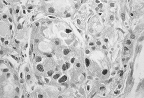
Fig. 6: Light micrograph of original thyroid before xenografting (original magnification 360 ×).
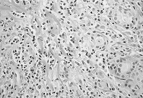
Fig. 7: Light micrograph of thyroid xenograft from patient in Fig. 6 in an NIH-3 mouse (original magnification 360 ×).
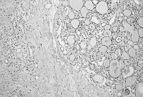
Fig. 8: Light micrograph of thyroid xenograft from patient in Fig. 6 in an SCID mouse. The original histologic study of the thyroid showed that mild infiltration of lymphocytes. Eight weeks after xenografting into mice, lymphocytic infiltration was more prominent in the NIH-3 mouse than in the SCID mouse (original magnification 360 ×).