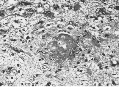
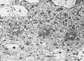
Fig. 5: (A) Light micrograph of a cerebral cortex section from a SHRSP in the 8-week treatment/withdrawal group, showing large cystic structures (C), extensive malacia (M) and fibrinoid necrosis in the vascular wall (large arrows). The bar represents 250 µm. (B) A higher magnification of Fig. 5A, showing a blood vessel with marked deposition of fibrin in the thickened wall (fibrinoid necrosis), surrounded by neuropil infiltrated by leukocytes and intense gliosis. The bar represents 50 µm.
[ Return to text ]