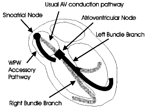
Fig 1. Diagrammatic representation of the heart showing the Wolff-Parkinson-White (WPW) accessory pathway and the normal atrioventricular conduction pathway.