Managing Colles' fractures in rural practice
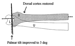
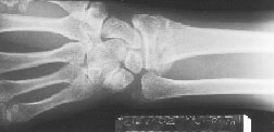
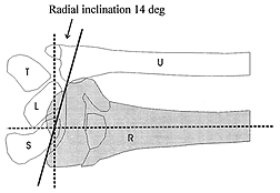
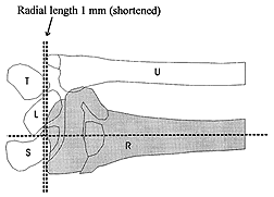
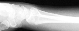
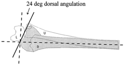
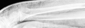
Fig. 1. Typical Colles' fracture before reduction in posteroanterior (top) and lateral (middle) views, and after reduction in the lateral view (bottom), showing the radius (R), ulna (U), scaphoid (S), lunate (L) and triquetral (T) bones.
[ Return to text ]