Managing Colles' fractures in rural practice
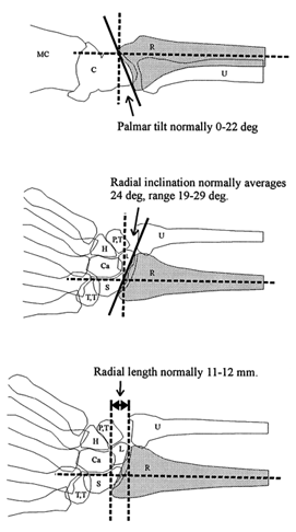
Fig. 4. Normal anatomy of the distal radius in lateral (top) and posteroanterior views (middle and bottom), showing the radius (R ), ulna (U), carpal bones (C), metacarpals (MC), trapezoid and trapezium (T,T), scaphoid (S), capitate (Ca), hamate (H), lunate (L), and pisiform and triquetral (P,T) bones.Ulnar styloid fractured through its base rather than more distallyHigh kinetic energy mechanism of injuryDevelopment of pain not relieved by recasting
[ Return to text ]