Volume 10: pp. 109–110

ccbr_vol10_weisman_bouton_spetch_wasserman_iconA Social History of the Founding of the Conference on Comparative Cognition and the Comparative Cognition Society
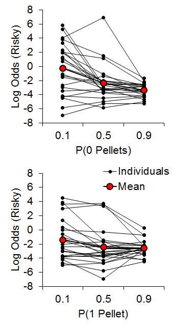
Reading Options:

Continue reading below, or:
Read/Download PDF| Add to Endnote

Ronald G. Weisman
Queen’s University

Mark E. Bouton
University of Vermont

Marcia L. Spetch
University of Alberta

Edward A. Wasserman
University of Iowa


This memoir is a brief history of the founding of the Conference on Comparative Cognition and the Comparative Cognition Society. The text represents the best recollections of the authors, who together founded the Conference. In the 1980s, Ron Weisman visited Melbourne, Florida, regularly to enjoy the warm weather in March and to visit friends at Florida Tech. Over time, he began thinking about sharing the Melbourne experience with other comparative cognition scientists. He discussed the idea with Mark Bouton, Marcia Spetch, and Ed Wasserman in the late 1980s: it is probably no accident that all four taught on campuses that experience harsh winters. By the early 1990s, the group began planning the meetings in earnest. Together, all four became known as the steering committee—or “steers” for short. The steering committee began meeting as a group and in pairs over the next few years to plan the conference. They decided on a name (the Conference on Comparative Cognition), and Ed Wasserman provided the acronym, CO3. Suzy Bouton did the wonderful logo. The committee discussed the lengths of the talks (5, 10, and 20 minutes). Mark Bouton suggested including very short, 5-minute talks, borrowed from the Winter Conference on the Neurobiology of Learning and Memory (Park City, Utah). At CO3, the 5-minute talks evolved into “spoken posters,” complete in themselves, practiced, polished, and informative. Graduate students were allowed to present these brief talks from the beginning, and faculty members were encouraged to do likewise. However, in practice, faculty give 10-minute talks, or, less commonly, 20-minute talks. Other more pragmatic, but important, decisions dealt with providing snacks and drinks at the meetings and when to schedule sessions.

Most important, the steering committee discussed what the meeting was going to be about. They decided that CO3 was to be about comparative cognition in the broadest sense, with encouragement to report on the standard laboratory species and on more naturally occurring species. By defining cognition broadly, they were able to avoid the squabbles then current between more behaviorally oriented and more cognitively oriented scientists.

CO3 first met March 17–20, 1994, at the Holiday Inn on the Ocean in Melbourne. Slightly fewer than three dozen scientists attended, but CO3 was off to a promising start. The attendees liked the meeting, and more than half said they would attend often if not every year. In those early years, more pelicans attended, and more alcohol was consumed.

Noise issues in the normal meeting rooms prompted the hotel to move us to a swank, oceanfront, two-story, glass-walled penthouse. For a time, the luxurious penthouse was perfect for meetings and great evening parties. But eventually rising room rates and noise from the Holiday Inn’s oceanfront entertainment drove CO3 a few miles north to the Hilton Hotel, which served CO3 well until the conference outgrew the limited, but excellent, accommodations. Conveniently, the Radisson Hotel directly up the beach was larger and a better business partner.

While the group met at the Hilton, in 1997, Suzanne MacDonald began to design and sell shirts commemorating the meetings. The designs always gracefully depicted an animal native to Florida. Suzanne joined the steering committee soon thereafter. That the meetings were held in warm, sunny Florida was always important. Mike Brown, later a member of the steering committee, gave the meeting its informal description: “It’s surf and science,” he said with a smile. Melbourne is a relatively quiet town and over the years Mark Bouton and then Marcia Spetch and Suzanne MacDonald sought out other venues; nevertheless, in direct comparisons, Melbourne always offered greater value than better-known towns. Cost has remained important because the meeting attracts so many graduate students.

In 1997, it became obvious that CO3 was growing into what we had all hoped it would become—a successful annual meeting of like-minded scientists. Just to be clear, CO3 was governed by an unelected steering committee, a sort of benign dictatorship. There was no plan for succession. The rapid growth in the number of presenters made it clear that the same few people could not be expected to continue to carry the responsibility, year after year, for a meeting projected to soon include 200+ scientists. At the request of Ed Wasserman, the steering committee met and discussed the next phase: incorporation as a scientific society, the Comparative Cognition Society. The overwhelming majority of presenters favored incorporation, but a few, including a member of the steering committee, did not. Ron Weisman responded to the majority opinion and carried through the incorporation in Florida, where the fees and responsibilities of running a nonprofit corporation are not onerous. We met as a society for the first time in 1999. By that time, we had assembled a rotating cadre of colleagues who worked hard to make sure that the meeting ran smoothly. After incorporation, we set up elected governance to take the Comparative Cognition Society forward into the new century.

Since incorporation, the Society has thrived. Members of the executive board (an expanded and elected version of the steering committee) have included both original members and new faces: Mike Brown, Bob Cook, Jon Crystal, Jeff Katz, Suzanne MacDonald, Marcia Spetch, Ed Wasserman, Ron Weisman, and Tom Zentall. Bob Cook has published two cyber books under the Comparative Cognition Society’s imprint. Ron Weisman and Bob Cook founded and took the first six-year turn editing the Society’s successful online journal, Comparative Cognition and Behavior Reviews. As first suggested by Ed Wasserman, leaders in the field of comparative cognition have been honored with a CCS Distinguished Researcher award, a Master Lecture, and a special issue of Behavioral Processes.

Over the years, CO3 has included talks on over 100 species by scientists from over a dozen countries. Through the political and economic crises of the early 21st century and the Society’s incorporation, attendance at CO3 has grown and remained strong. We are pleased to report that each year brings both returning and new researchers, together with students ready to become the future of our field.

Volume 10: pp. 107

In Memory of Ronald G. Weisman

(September 14, 1937 – January 27, 2015)

Ron Weisman

Reading Options:

Continue reading below, or:
Read/Download PDF| Add to Endnote

Christopher B. Sturdy & Marcia L. Spetch
University of Alberta

With his family by his side, Ron Weisman, Professor Emeritus, Departments of Psychology and Biology, Queen’s University, died at home on 27 January 2015. Ron obtained his Ph.D. from Michigan State University in 1964 and was hired as Assistant Professor of Psychology at Queen’s in that same year. Ron was promoted to Associate Professor in 1970, Professor in 1977, cross appointed to the Department of Biology in 1993, and finally promoted to Professor Emeritus in 2000. In sum, Ron was a professor at Queen’s for over 50 years. He is well known for his numerous significant contributions to our understanding of animal learning, cognition, and behaviour. Maybe more important, but not so easily tallied with facts and numbers, are the more qualitative and impactful contributions that Ron made to the research areas in which he was so totally and passionately invested during his long and productive career but that escape the accountant’s ledger. Of these less quantifiable, but absolutely important contributions, one cannot hope to produce a comprehensive report here. And Ron himself would not want such a thing. “Too many words that no one is likely to read or care about” would probably be his quip in response to such an idea. No, the manner in which Ron operated and conducted himself is best described using the words of those who have commented about Ron’s influence in the days since his passing. Strong themes like “force of nature”, “intellectually challenging”, “passionate”, “inspiring” are a constant in Ron’s colleagues’ narratives shared in conversations, social media, and emails. Never one to back down from a challenge, Ron reinvented his research career from the ground up when he realized an opportunity to pursue new more challenging but meaningful problems. This categorical change came when Ron was at a point in his career in which most people would be happy to simply maintain the currently successful status quo until retirement. Not Ron. Instead, and in spite of, or perhaps, because of, the fear of the unknown, Ron forged a second, even more well-known career for himself, combining research in learning, cognition, ethology, and neuroscience in a manner not often done, certainly not with the same effect. While on this new path, Ron continued to make significant contributions to the scientific literature and to the field through the founding of the Comparative Cognition Society, and their flagship online and open access journal, Comparative Cognition & Behavior Reviews. Perhaps Ron’s most enduring legacy will be of the contributions that he made to the mentorship and encouragement of young scientists. Many successful scientists owe their “academic legs” to Ron’s strong and generous support and wisdom. Ron posed challenging questions and championed points of view that were sometimes controversial and always aimed at pushing back the darkness to, as Ron put it, “Explain nature”. Ron always managed to be engaging, encouraging, and able to coax the absolute best out of everyone who was willing to meet his enthusiasm and level of commitment to science. Ron’s enthusiasm, wit, candor, compassion, and his huge smile will be sorely missed by all who had the pleasure of knowing him. What a guy.

Volume 10: pp. 73–105


ccbr_vol10_qadri_cook_iconExperimental Divergences in the Visual Cognition of Birds and Mammals

Muhammad A. J. Qadri & Robert G. Cook
Department of Psychology, Tufts University

Reading Options:

Continue reading below, or:
Read/Download PDF | Add to Endnote


Abstract

The comparative analysis of visual cognition across classes of animals yields important information regarding underlying cognitive and neural mechanisms involved with this foundational aspect of behavior. Birds, and pigeons specifically, have been an important source and model for this comparison, especially in relation to mammals. During these investigations, an extensive number of experiments have found divergent results in how pigeons and humans process visual information. Four areas of these divergences are collected, reviewed, and analyzed. We examine the potential contribution and limitations of experimental, spatial, and attentional factors in the interpretation of these findings and their implications for mechanisms of visual cognition in birds and mammals. Recommendations are made to help advance these comparisons in service of understanding the general principles by which different classes and species generate representations of the visual world.

Keywords: Pigeons; Humans; Visual cognition; Spatial attention; Perceptual grouping; Perceptual completion; Visual illusions

Corresponding author:
Dr. Robert G. Cook Department of Psychology; Tufts University 490 Boston Ave.; Medford, MA 02155, USA; Phone: 617 627-2456, Fax: 617 627-3181; E-mail: Robert.Cook@tufts.edu

This research was supported by National Eye Institute grant EY022655.


Visual cognition is critical to the behavior of complex animals. It generates the working internal cognitive representations of the external world that guide action, orientation, and navigation. The extensive study of the human animal has dominated the theoretical and empirical investigations of vision and visual cognition (Palmer, 1999). In comparison, the psychological investigation of visual cognition in other animals has received far less attention. Not surprisingly, the examinations of nonhuman primates have been of most interest precisely because their visual system most closely resembles our own. Despite this focus on primates, there is a long and distinguished record of comparative research with non-primate species that has profoundly enhanced our understanding of vision and its underlying mechanisms (e.g., Hartline & Ratliff, 1957; Hubel & Wiesel, 1962; Lettvin, Maturana, McCulloch, & Pitts, 1959; Reichardt, 1987). An appreciation of the entire spectrum of visually driven cognitive systems and how vision is implemented in different nervous systems is key to a complete and general understanding of the evolution, operations, and functions of vision and its role in cognition and intelligent behavior (Cook, 2001; Cook, Qadri, & Keller, in press; Lazareva, Shimizu, & Wasserman, 2012; Marr, 1982).

One of the most fruitful investigations of these comparative questions has focused on the visual behavior of birds, especially in comparison to mammals (Cook, 2000, 2001; Zeigler & Bischof, 1993). There is no question of the importance of the visual modality for these highly mobile creatures. Beginning with their origins within the lineage of feathered theropod dinosaurs (Alonso, Milner, Ketcham, Cookson, & Rowe, 2004; Corwin, 2010; Lautenschlager, Witmer, Altangerel, & Rayfield, 2013; Sereno, 1999), birds have subsequently and rapidly evolved on a number of fronts, including pulmonary physiology, the development of endothermy, distinctive strategies for reproduction and growth, and their central neuroanatomy (Balanoff, Bever, & Norell, 2014; Xu et al., 2014). Over that time, birds have evolved central and visual systems that are well suited for high-speed flight within the restrictions of muscle-powered transport. While quite large relative to birds’ body size, the avian brain is still small compared to primates’ in absolute size. Given the computational complexity and problems associated with vision, the difficulties of building flexible and accurate optically based machine vision systems, and the considerable and large portions of the primate brain devoted to visual cognition, the small size and visual excellence of the avian brain presents an interesting challenge and scientific opportunity. Given their high visual functionality and small absolute brain size, birds provide an excellent model system for guiding the practical and efficient engineering of small visual prostheses, while simultaneously advancing our general theoretical understanding of visual cognition.

The ancestors of modern-day birds and mammals followed contrasting diurnal and nocturnal evolutionarily pathways during the Mesozoic era, and as a result, these two major classes of vertebrate have evolved to rely more heavily on structurally different portions of their nervous systems to mediate visually guided behavior (Cook et al., in press). Most likely because of their nocturnal origins, mammals have evolved solutions to the challenges of vision that developed into numerous lemnothalamic cortical mechanisms and areas that primarily mediate visual cognition (Felleman & Van Essen, 1991; Homman-Ludiye & Bourne, 2014; Kaas, 2013). In contrast, birds use a collothalamically dominant vision system, mediated by the tectum and related structures, to process visual information. From one perspective, birds may represent the evolutionary zenith of the animals that rely on this ancient primary ascending pathway for vision. The complementary pathway present in both animal classes, however, is still critical to visual function. The collothalamic pathway involving the superior colliculus and pulvinar have well established and important visual and attentional functions in mammals (Müller, Philiastides, & Newsome, 2005; Petersen, Robinson, & Morris, 1987; Robinson, 1972), while the visual Wulst in the avian lemnothalamic pathway may play similar roles in birds (Shimizu & Hodos, 1989). Given these differences in the relative weighting and possible functions of these different pathways for each class, the direct comparison of these two types of vision systems provides theoretically revealing comparative information regarding the implementation and role of general, specific, and alternative routes to representing and understanding the visual world (Marr, 1982).

Pigeons have been the dominant avian model and focus species for this comparison. Years of intensive study have resulted in this bird’s visual, cognitive, and neural systems being the best understood of any avian species (Cook, 2001; Honig & Fetterman, 1992; Spetch & Friedman, 2006a; Zeigler & Bischof, 1993). Because the study of visual cognition in mammals has been dominated by studies of humans, the outcomes of the laboratory studies of pigeons have naturally and frequently been compared with our own visual behavior. More important, the extensive theoretical concepts developed from research on human visual cognition have regularly served as a guide for developing investigations of avian visual cognition. Combined, these forces have produced an extensive number of studies in which these two contrasting vertebrate species have been tested with identical or highly similar visual stimuli.

What is the current status of this scientific comparison of pigeon and human visual cognition? Moreover, what similarities and differences have been established regarding how these different classes of animals solve the challenging problems of visually navigating and acting in an object-filled world? On one front, a number of similarities have been established. For example, humans and pigeons discriminate letters of the English alphabet in highly analogous ways, suggesting that shape processing across these species may share similarities (D. S. Blough, 1982; D. S. Blough & Blough, 1997). Looking more deeply at the mechanisms underlying such findings, the early processes responsible for dimensional grouping appear to share similar organizational principles, with their combination, use, and recognition of color, shape, and relative illumination operating in ways that appear comparable (Cook, 1992a, 1992b, 1993; Cook, Cavoto, Katz, & Cavoto, 1997; Cook, Cavoto, & Cavoto, 1996; Cook & Hagmann, 2012; Cook, Qadri, Kieres, & Commons-Miller, 2012). The investigation of visual search behavior has suggested that the search for targets in noise is governed by the same basic parameters across species (D. S. Blough, 1977, 1990, 1992, 1993; P. M. Blough, 1984, 1989; Cook & Qadri, 2013). Extensive research examining the pictorial discrimination of various objects derived from “geons” has suggested that pigeons and humans share commonalities in their processing of these stimuli as well (Kirkpatrick-Steger, Wasserman, & Biederman, 1996, 1998; Van Hamme, Wasserman, & Biederman, 1992; Wasserman & Biederman, 2012; Wasserman, Kirkpatrick-Steger, Van Hamme, & Biederman, 1993; Young, Peissig, Wasserman, & Biederman, 2001). These different parallels carry the important theoretical implication that the natural structure of the visual world may restrict the classes of computational solutions to a fairly small set of mechanisms, even across quite different biological visual systems. Thus, whether an animal is using a collothalamic- (birds) or lemnothalamic-dominant (mammals) visual system, they may operate using similar computational and processing principles because of the structure of the visual world (J. J. Gibson, 1979; Marr, 1982).

Despite the existence of these numerous experimental parallels, this “representational equivalence” hypothesis is surely not a comprehensive description. One might reasonably question given just the simple disparity in absolute size and internal organization of the brains of birds and mammals how these visual systems could function comparably. All of our additional cortical areas and tissue must allow us some enhanced functionality, such as mental imagery or manipulation. Consistent with this line of thinking, a number of experimental findings create problems for such a “representational equivalence” hypothesis. These findings include discrepancies, anomalies, or divergences in the apparent perceptual behaviors of these two species across many experiments. These divergences have not been just one or two isolated occurrences in a few limited settings, which may be overlooked, brushed aside, or dismissed. To the contrary, many occur in persistent clusters in theoretically relevant areas. The purpose of this article is to collect and evaluate lines of these divergent findings and their implications for theories of comparative visual cognition.

Conceptual Overview and Framework

There are a number of areas in which divergent findings have been reported involving pigeons and humans. Examining such divergences is an important way to evaluate the scope and limitations of representational equivalence and to identify potential functional differences. Precisely identifying and isolating where avian and mammalian visual systems differ and where they share commonalities is crucial to reverse engineering their computational mechanisms and evaluating all of the different possible alternative routes to visual representation.

The quintessential outcome of any one of these studies is that the perceptual responses of the pigeons fail to mimic those of humans (or vice versa depending on your taxonomic affection). For example, a number of psychophysical investigations have found that pigeons have poorer acuity and motion thresholds, lower flicker fusion thresholds, and differences in their processing of color relative to humans (Bischof, Reid, Wylie, & Spetch, 1999; P. M. Blough, 1971; Hendricks, 1966; Wright & Cumming, 1971). Beyond these differences in basic visual sensory function between pigeon and human, another difference is the pigeon’s strong propensity to attend to smaller local features or portions of stimuli rather than grasp the larger global form (Aust & Huber, 2001, 2003; K. K. Cavoto & Cook, 2001; Cerella, 1977, 1986; Emmerton & Renner, 2009; Kelly, Bischof, Wong-Wylie, & Spetch, 2001; Lea, Goto, Osthaus, & Ryan, 2006; Navon, 1977, 1981; Vallortigara, 2006). This same local precedence has also been evidenced to a certain degree outside of the operant chamber, during spatial cognition investigations of landmark use (Kelly, Spetch, & Heth, 1998; Spetch, 1995; Spetch & Edwards, 1988). While pigeons are able in the right circumstances to process global information, processing information at larger spatial scales seems far more difficult for them (Cook, Goto, & Brooks, 2005; Fremouw, Herbranson, & Shimp, 2002; Kelly et al., 2001). Such psychophysical and attentional differences are noteworthy and significant and likely play important roles in resolving some of the findings considered in more detail below.

To make this review manageable, however, we have restricted our considerations to four topics that have generated a larger corpus of divergent findings in the domain of visual cognition. Specifically, we look at the discrimination of different arrangements of line-based shapes, the grouping and integration of dot-based perceptual information, the perceptual completion of spatially separated information, and the perception of geometric visual illusions. These are selected because they each represent persistent areas of experimental divergences that have centered on processes that are fundamental to visual cognition theoretically.

The pivotal issue in each case centers around whether any dissimilarities between avian and primate perception reflect a true qualitative difference in how the two classes of animals visually perceive and process the world or instead reflect experimental artifacts or limitations that do not represent the true, underlying state of affairs. Despite the best intentions of the experimenters to nominally investigate the same question across these species by testing similar or identical stimuli, many different variables and procedural issues could potentially produce a given divergent result.

Some of these complicating issues may be related to the experimental or discriminative procedures involved with testing different species. Differences in visual angle, subject training, previous experience, or experimental instructions are all examples of this type of issue. For instance, humans are often explicitly instructed about what features are relevant during testing. In marked contrast, pigeons always have to discover the relevant visual features on their own based on differential reinforcement. Consequently, the two species may ultimately perceive or attend to different features or aspects of superficially identical displays. If the latter occurs, this naturally limits any implications for our deeper understanding of visual processing. To draw the strongest conclusions, both species must attend to the same features in the experimental displays.

Likewise, discrepancies may stem from other attentional or cognitive differences between these species. As mentioned, pigeons regularly exhibit a bias to attend to spatially local information in preference to more globally available information in visual discriminations. Humans contrastingly appear to be much more global in their allocation of attention. Because of its potential impact, a framework for thinking about how animals might spatially attend to stimuli is worth considering at this juncture.

Figure 1 shows two important facets of spatially controlled attention. The first is the size of the area processed in a single visual scan. This might be best thought of as a visual “aperture,” an adjustable area, presumably circular, over which information is processed without any additional eye fixations. The second important component is the size of the spatial search area that is analyzed or integrated over using a series of successive fixations of this visual aperture around the display. This is also adjustable, and presumably operates over a larger region to integrate information. This search area could be as expansive as the whole operant chamber, or it could be limited to the regions of the display critical for correct discrimination. Both of these spatial components, search aperture and area, likely can vary independently, although a trade-off necessarily occurs between them. When a small visual aperture is employed, for example, greater scanning over a display may be strongly encouraged.

Figure 1. Depiction of the different hypothesized modes of spatial attention mechanisms and their critical features. We propose that pigeons use an attentional aperture over areas of the display to visually process information in the operant chamber. These distinctions yield two distinct types of global strategies: sequential integration and global perception. As depicted by the pigeon on the bottom, sequential integration applies a small attentional aperture to multiple spatial locations, integrating the information from each aperture to yield a global percept. The pigeon on the right depicts global perception, where a global feature is extracted from a single, large aperture. In contrast, the pigeon on the left applies a small aperture to only a single location, exemplifying a mode of (spatially) restricted local processing.

Figure 1. Depiction of the different hypothesized modes of spatial attention mechanisms and their critical features. We propose that pigeons use an attentional aperture over areas of the display to visually process information in the operant chamber. These distinctions yield two distinct types of global strategies: sequential integration and global perception. As depicted by the pigeon on the bottom, sequential integration applies a small attentional aperture to multiple spatial locations, integrating the information from each aperture to yield a global percept. The pigeon on the right depicts global perception, where a global feature is extracted from a single, large aperture. In contrast, the pigeon on the left applies a small aperture to only a single location, exemplifying a mode of (spatially) restricted local processing.

The combination of these two attentional attributes results in four modes by which information can be extracted from a visual display. One means of globally processing information over a spatial area would be to employ a large visual aperture and reduce the need for much successive scanning. This is much like what occurs in parallel search or perceptual grouping, especially in humans, where information is extracted or discriminated rapidly over a large spatial extent. A second way is to employ a smaller or more local visual aperture with numerous successive scans of a display that are then combined in later computations. This would yield a process similar to serial visual search and is a different way that animals could successively integrate information over a spatial extent. We think it is important to distinguish between these alternatives when thinking about their impact on global processing. We use the term global perception to indicate the use of a single, large spatial aperture, while the term sequential integration will be used to indicate the hybrid use of a smaller local aperture with a global scanning and integrating strategy. The third approach would be to use a smaller visual aperture with a spatially restricted scanning strategy, without integrating information from separate scans. This would lead to a more particulate perception of the display. We will use the term restricted local processing to describe this attentional approach, and to distinguish it from the sequential integration strategy that may have a similarly sized aperture, but a more expansive scanning strategy. The logical fourth alternative is one that combines a large visual aperture distributed widely over a large spatial area employing a large number of scans or fixations. This last method has similarities to how we experience and navigate the natural world. Given the restricted spatial scale of the typical operant setting, we think this mode plays a less prominent role in the findings below (but merits considerable more research attention). We think these different processing distinctions are worth keeping in mind when evaluating the results collected here.

Line-Based Shape Processing

Overall, the review is divided into four sections covering each broad topic area, followed by a discussion that integrates the interim conclusions of each section in service of answering the larger theoretical question of how avian and mammalian visual cognition are similar and different. This first section examines divergent findings involved with the processing of shape discriminations by pigeons. Because the motivations, stimuli, and tasks are different from each other, they do not easily form a shared theoretical focus. Thus, the possibility that we are combining different underlying phenomena by grouping them should be held in mind. They do share, perhaps importantly, the common feature of using stimuli comprised of different complex arrangements of line segments.

Stimulus Configuration

One important visual outcome in humans is a set of findings classified as configural superiority effects. Here, humans perform better when the arrangement or context of simple elements create configural or emergent properties that facilitate discrimination. The important theoretical idea captured by such results is that the emergent or holistic features of some stimuli precede or dominate the processing of their component elements (Kimchi & Bloch, 1998; Pomerantz, 2003; Pomerantz & Pristach, 1989).

The classic example involves a simple discrimination of the diagonal tilt of two lines, as shown in Figure 2A. With the addition of a redundant “L” context to the tilted lines, this transforms into a discrimination of a “triangle” versus an “arrow,” facilitating performance in people (Pomerantz, Sager, & Stoever, 1977). Because of its importance to the visual mechanisms of holistic and analytic processing, this same type of visual phenomenon has been examined in pigeons.

D.S. Blough (1984) reported the first results testing configural-like stimuli with pigeons. He found mixed results in the two experiments briefly described in that chapter. Using a simultaneous discrimination and three highly experienced birds familiar with making letter discriminations, he reported the results of a discrimination with pattern-producing configural contexts. These consisted of a contextual L that produced an emergent “triangle” or “arrow” for rightward or leftward diagonal lines, or a “U” or “sideways U” as added to horizontal and vertical lines. Through a series of reacquisitions, the configural patterns were learned more quickly and responded to faster than the context-free line discriminations, suggesting that pigeons experience the same kind of configural superiority effect as humans. Subsequent investigations of this type of effect were not as encouraging, however.

In the same chapter, Blough also reported tests with line stimuli in which the added context resulted in a figure that formed a possible 3D object, as well as equally complex contexts that had no obvious 3D interpretation (see Figure 16.4 in Blough, 1984 for examples). For humans, the configural 3D figures were far easier to discriminate because they formed different global objects. For pigeons, this form of configural discrimination was reported as difficult to learn, regardless of the potential 3D interpretation. This suggests that the different line placements did not produce configural depth relations or objects in pigeons, or if they did, the resulting figures did not aid in the discriminability of the display.

In a more extensive investigation of this general issue with a larger number of pigeons, Donis and Heinemann (1993) also trained their animals to discriminate between rightward and leftward sloped diagonal lines in isolation or with the same addition of an L context (i.e., the classic “triangle” vs. “arrow”). In Donis and Heinemann’s study, however, the pigeons showed reduced accuracy when the discriminated lines were embedded in the configural L context. Unlike humans, the pigeons were more accurate when discriminating the lines in isolation. There are notable methodological differences that could have contributed to this difference from Blough’s brief description. Whereas the pigeons in D.S. Blough (1984) pecked directly at the correct stimulus, the pigeons in Donis and Heinemann had a subsequent spatial choice after pecking the discriminative stimulus. Also, the stimuli in Donis and Heinemann’s investigation were about four times larger than those in D.S. Blough (1984), raising the possibility that limits of processing might be related to the size of the stimuli. This latter possibility would suggest that Blough’s pigeons may have employed a global processing strategy, while Donis and Heinemann’s pigeons could have relied on sequential integration or restricted local processing. Donis and Heinemann’s results are not unique, however.

Kelly and Cook (2003) conducted three experiments with different groups of pigeons, examining the role of contextual information on the discrimination of diagonal lines and a mirror-reversed L discrimination. One group was tested in a target localization task using texture displays made from either repeated lines or configural patterns. The second group of pigeons was tested in an oddity-based same/different task. Similar to Donis and Heinemann (1993), the pigeons exhibited a reduction in target localization or same/different choice accuracy with the pattern-producing stimuli in comparison to the simple discrimination of diagonal lines. This was true across presentations using both small and large sizes of the stimuli, suggesting that visual angle was not particularly important. Furthermore, a second pair of configural stimuli involving a “positional discrimination” was also tested. Here the “featural” stimuli consisted of an L versus a mirror-reversed L, and the redundant context that permitted emergent features was a diagonal line (see Figure 1B in Kelly and Cook, 2003, for examples of these stimuli). This type of stimulus also showed no differences compared to the elemental and configural conditions in the same/different task, although it did reveal a configural superiority effect in the target localization task. This configural superiority effect may have been caused by the high similarity of the textured regions produced by the mirror-reversed elements. Nevertheless, in general, the pigeons in this study were typically better when the discriminative line stimuli were presented in isolation than in a configural organization.

A different configural effect found with humans involves stimuli using component line elements arranged to form a human face. Testing stimuli that produce a face superiority effect in humans, Donis, Chase, and Heinemann (2005) found that their pigeons’ capacity to discriminate the feature of a U (the ”smile”) versus its flipped counterpart ∩ (the “frown”) was impaired when a triplet of dots arranged as “eyes” and a “nose” was placed above it. The pigeons were further impaired when these features were enclosed within a larger ellipse, a condition in which humans see a clear and readily discriminable face. Thus, instead of promoting discrimination with the addition of these configuration-producing elements, for the pigeons, these additional features obscured the critical portion of the discrimination. Given that the pigeons failed to learn to discriminate these complete, configural displays even with extensive training, these authors suggested that the additional context increased the similarity between the configural stimuli for the pigeons instead of accentuating or producing new featural differences as it appears to in humans.

Thus, despite the promising start, the preponderance of the evidence suggests that pigeons tend not to exhibit the same configural superiority effects as observed in humans. As accessed by several experiments using different discrimination approaches, pigeons do not consistently benefit from the addition of contextual or configural information in these stimuli that humans find beneficial. Instead, the more typical result seems to be that the pigeons exhibit a form of configural inferiority effect, where the added elements interfere with discrimination by increasing the similarity of figures. This suggests that the two species are deriving or attending to different features within these stimuli.

Search Asymmetry

Another line of divergent research in this area is related to visual search asymmetries. In multiple investigations with humans, Treisman and various colleagues have found that not all sets of features produce identical modes of visual search (Treisman & Gormican, 1988; Treisman & Souther, 1985). In particular, Treisman and Souther found that some shapes could produce parallel search (like a single Q embedded in Os; see Figure 2B), while a reversal of the same features would result in serial-like search (a single O embedded in Qs). Treisman’s theoretical analysis of these search asymmetries focused on the fact that distinctive visual features were selectively activated for one type of search but not the other, such as detecting the presence of the singular line when a Q was the target in a set of Os.

Because of its theoretically revealing nature, Allan and Blough (1989) examined visual search in pigeons with variations of two types of search asymmetry stimuli previously established with humans. Their search displays included the search between O and Q and between triangles with and without a gap along one edge. Overall, they found no search differences depending on which line shapes were the target or distractors; the presence or absence of a feature in the target generally did not affect their search speed or accuracy for either the added line or gap stimuli. This divergence from humans—the apparent absence of a feature search ­asymmetry—is likely not due to pigeons being unable to exhibit such asymmetries in search tasks. Using search displays made up of groups of smaller squares of differing colors, Pearce and George (2003) found that pigeons did show asymmetries in accuracy when distinctive color features are located in the target rather than the distractors. This suggests that the divergence between humans and pigeons found by Allan and Blough may be specifically tied to the dimensional or featural processing of lines or shapes.

In later work, D. S. Blough (1993) examined the use of cues in stimuli that were square-like and contained an additional line and/or gap. On any given trial, an array of 32 stimuli in four rows was displayed, and the pigeon had to identify which stimulus within the array was unique. In this visual search task, Blough analyzed how reaction time varied according to the specific stimulus-pairs tested. He found that the pigeons appeared to independently attend to the presence of the additional lines in the different stimuli and to either location at the top or bottom of the shapes. The gap in the stimulus appeared not to capture attention, consistent with Allan and Blough’s (1989) earlier results. Importantly, the consistent and measurable within-stimulus effect highlights how even small differences in spatial attention directed toward different local areas of stimuli may be a potentially important concern in the analysis of stimuli.

Vertices and edges

For humans, one important outcome of studying visual cognition is our reliance on information at the junctions or vertices of objects for their recognition. Biederman (1987), for example, has found that the equivalent deletion of the vertices of an object is far more detrimental to its subsequent recognition by humans than the deletion of contour information in the middle of line segments (see Figure 2C). The analysis and prioritization of vertices as a critical feature also plays a classic and prominent role in object recognition algorithms by computers (e.g., Harris & Stephens, 1988; Trajković & Hedley, 1998). One possible reason for this reliance is that junctions contain greater information content to aid in deriving the non-accidental structural relations of an object’s surfaces as compared to edges. Focusing on vertices thus reduces the probability of accidental visual properties causing misperception of objects. It is natural to extend this question to whether pigeons use this feature in the recognition of objects.

Figure 2. Examples of stimuli from different experiments focused on line-based figural processing. Panel A depicts a classic human configural superiority effect that has been tested with pigeons. Panel B shows a search asymmetry task in which the unique element contains an added line, which benefits humans in searching for the target, but not pigeons. Panel C depicts two-dimensional shape stimuli that have had either vertices/cotermination or line segments or edges removed.

Figure 2. Examples of stimuli from different experiments focused on line-based figural processing. Panel A depicts a classic human configural superiority effect that has been tested with pigeons. Panel B shows a search asymmetry task in which the unique element contains an added line, which benefits humans in searching for the target, but not pigeons. Panel C depicts two-dimensional shape stimuli that have had either vertices/cotermination or line segments or edges removed.

Several investigations have suggested that pigeons might differ from humans in this regard. The earliest, most prominent, and most complete example was reported by Rilling, De Marse, and La Claire (1993). They trained pigeons to discriminate different shapes using both 2D (outlined square vs. triangle) and 3D (outlined cube vs. prism) figures. They then tested the pigeons by deleting different portions of the shapes at either the center of the lines or at their junctions. Across both sets of stimuli, the pigeons were more disrupted by the elimination of the line segments midway between the vertices than at the vertices. This outcome contrasts markedly with the typical human finding.

Several unpublished observations have since been consistent with Rilling et al.’s (1993) findings. An unpublished experiment from the Comparative Cognition Laboratory at the University of Iowa (Wasserman, personal communication) further suggested that vertices made less of a contribution to the recognition of geons by pigeons. Here segments were removed from complex line drawings of objects at the vertices and between the vertices. Similar to Rilling et al, the pigeons performed better in the former conditions. This outcome was complicated by additional differences between the conditions, however, as it was difficult to equate the length of the remaining line segments between these different conditions because of their complexity. In our own lab, we have found something similar in a target visual search task using texture stimuli. Cook (1993) reported that linear arrangements of distractors were more interfering than either randomized or spaced distractors in such a task (see examples of these stimuli in Figure 14.1 of Cook, 1993, p. 247). In subsequent unreported experiments, we found that edge-like linear distractors interfered more with target search than distractors made from the same number of elements but forming vertex-like right angles. This outcome hints that the edges of the square target regions were more critical to their identification by the pigeons than the corners.

Using a different approach, Peissig, Young, Wasserman, and Biederman (2005) examined how similarly pigeons process shaded complex objects and line drawings of the same objects. Using a variety of training and transfer designs, it appeared that the pigeons did not use the common edges or edge relations across shaded and line objects to mediate their discrimination, as their transfer was poor between these sets of stimuli. The results suggest instead that the pigeons used different representations of each group of stimuli, perhaps based on the availability of surface characteristics. Peissig et al. suggested that their pigeons may have placed greater importance on surface features than edges in learning these discriminations. Not all studies have found this type of result.

In contrast, a recent experiment by Gibson, Lazareva, Gosselin, Schyns, and Wasserman (2007) suggested instead that line co-termination in complex stimuli is an important factor for pigeons. In their experiment, pigeons were trained to discriminate four shaded geons in a choice task. They used a “bubbles” technique, where differing amounts of information were randomly visible through a set of Gaussian apertures placed over the stimuli. Accuracy was averaged across these varying amounts and locations of visible information to identify which portions of the images were most important to the pigeons’ discrimination. Statistical pixel-based analyses of the resulting classification images indicated that both pigeons and humans used pixels near vertices more than edges or surfaces. Visual inspection of the classification images for the individual pigeons, however, do suggest that line segment, edge, and surface information may have also made important contributions to each bird’s idiosyncratic solution. Nonetheless, this work provides the best evidence yet that the vertices or co-terminations of the objects carry more weight than edges for the pigeons.

Taken all together, these sundry lines of investigation based on various aspects of processing line-based stimuli suggest that pigeons do not always readily reproduce the same visual phenomena observed in humans. Pigeons frequently show configural effects that are different from or possibly opposite those in humans. Pigeons appear not to exhibit the same feature-based search asymmetries as humans with seemingly comparable shape-based stimuli. Finally, pigeons do not always consistently prioritize junctions and co-terminations in shape discriminations like humans. While each of these conclusions involves a different type of discrimination, the one common feature across these investigations is the discrimination of simple lines and their relations. As outlined previously, the key question is whether these findings are truly capturing a difference in processing or represent some kind of experimental by-product or artifact.

One possibility is that any emergent structures from simple lines have a greater meaningfulness to humans. Perhaps these more impoverished stimuli require some abstraction to recognize their relation or correspondence to real objects. Humans may have this capacity, but the pigeon visual system may require more complete and realistic stimuli to properly process elements and their configurations (B. R. Cavoto & Cook, 2006). One reason that Gibson et al.’s (2007) results might differ, for instance, is that their object stimuli were more complete and realistic because of the presence of surface shading information. Furthermore, in humans, these types of stimuli can take advantage of linguistic labels or our greater experience at reading with complex line shapes. Each of these experiential factors could provide an advantage in processing lines and their configurations. This line of reasoning would suggest that examining arrangements that are more naturally salient to pigeons (such as those related to food or mating) could reveal processing more similar to that in humans.

Alternatively, pigeons and humans could have simply focused on different aspects of the stimuli because of experiential, instructional, or other cognitive differences. In humans, global perception of the stimuli allows all of the display’s features to create new unitary forms that are easy to discriminate. This might not be the case for pigeons, where local details of the stimuli might dominate their perception. There are several ways that this difference could have manifested in these experiments. One possibility is the use of experimental conditions that do not promote the use of larger visual apertures or global perception strategies by the pigeons. This stems from the fact that the majority of the pigeon studies varied neither stimulus size nor stimulus location during training. Fixed-size and fixed-position procedures are likely the best conditions for supporting restricted local processing strategies. Additional concerns in the same vein can also be raised regarding the differences in visual angle of the stimuli experienced by both species. Furthermore, humans often receive explicit instructions as to what to attend to, whereas the pigeons do not. Thus, before concluding the theoretical question of whether pigeons visually process line information or employ features in shape processing differently than humans, it would be valuable to consider and alleviate the possibility that the results are artifacts of restricted attentional strategies, stimulus size, or instruction.

If we ignore these concerns for a moment, the pattern of results raises the possibility that humans and pigeons process line-based visual features in different fashions. The machine vision literature is replete with different ways to combine the visual features corresponding to the edges and vertices of objects, as well as other higher-level shape features, to generate representations of objects in space. If there are differences in the way these line-based shape features are processed, despite the apparent similarities in the behavior of pigeons and humans in many settings, it would give rise to questions about how such features coalesce into representations that produce similar actions (Pomerantz, 2003). This larger issue is returned to subsequently in the general discussion.

Dot-Based Perceptual Grouping

Moving beyond the “simpler” line stimuli of the first section, the next area examines more complex stimuli perhaps best placed under the heading of perceptual grouping. Broadly conceived, perceptual grouping involves grouping identical, disconnected local elements into larger, global configurations. Some investigations of grouping using humans and pigeons have shown similar or overlapping patterns of responding. For example, as investigated by texture segregation, the early perceptual grouping of color and shape has generally been found to be similar across the two species (Cook, 1992b, 1992c, 2000; Cook et al., 1997; Cook et al., 1996; Cook, Katz, & Blaisdell, 2012). Based on such evidence, we have suggested that early vision is organized along highly similar lines, perhaps because of the importance of determining the extent and relations of object surfaces. Nevertheless, there are several areas where pigeons have consistently deviated from humans with stimuli that presumably involve similar grouping processes. A place to start is with larger global stimuli built from localized smaller dots.

Glass Patterns

In an important study in this area, Kelly et al. (2001) examined how pigeons and humans process Glass patterns. Glass patterns are theoretically revealing stimuli created by taking randomly placed dots, offsetting them appropriately, and superimposing the transposed result on the original stimulus (Glass, 1969; see Figure 3A). Humans readily perceive the global organization of the resultant Glass patterns from these dotted dipoles. Furthermore, humans detect circular or radial Glass patterns through random noise more easily than either translational or spiral patterns (Kelly et al., 2001; Wilson & Wilkinson, 1998). A similar sensitivity to circular information has been found with gratings in nonhuman primates (Gallant, Braun, & Van Essen, 1993). It has been hypothesized that this particular pattern superiority is caused by specialized concentric form detectors that might be the precursors to cortical face processing (Wilson, Wilkinson, & Asaad, 1997).

Using these types of dotted displays, Kelly et al. (2001) trained pigeons to discriminate vertical, horizontal, circular, and radial Glass patterns from a comparable number of randomly placed dots. Besides being generally more difficult for the pigeons, they found no differences in accuracy across the different global patterns regardless of their organization. When different numbers of the dots were placed randomly, creating noise in the displays, the pigeons continued to exhibit equivalent performance among the display types, unlike humans who showed the typical benefits of circular-like patterns. Kelly et al. suggested that this difference between species was potentially driven by the neurological differences between avian and primate visual systems. Consistent with this analysis, we recently extended these findings to a new species of birds, starlings (Qadri & Cook, 2014). Using Glass patterns that duplicated those tested with pigeons, the starlings’ behavior was highly similar to the pigeons’ with no differences found among the patterns.

Biological Motion

Another important area of visual cognition research involves the study of biological motion (Johansson, 1973). Coordinated moving points or dots that correspond to the motions of different articulated behaviors, such as in point-light displays (PLDs), powerfully invoke the perception of acting agents in humans (see Figure 3B). Humans can recognize a variety of actions and socially relevant features (e.g., age, gender, emotion) from these coordinated motions (Blake & Shiffrar, 2007). As a result, the study of action in humans has historically relied on these form-impoverished displays because they isolate motion-related contributions to action recognition. Because of their dominance in the study of action in humans, attempts have been made to examine action recognition in animals using PLDs with varying degrees of success (Blake, 1993; J. Brown, Kaplan, Rogers, & Vallortigara, 2010; Oram & Perrett, 1994; Parron, Deruelle, & Fagot, 2007; Puce & Perrett, 2003; Tomonaga, 2001). These include several investigations testing birds (Dittrich, Lea, Barrett, & Gurr, 1998; Regolin, Tommasi, & Vallortigara, 2000; Troje & Aust, 2013; Vallortigara, Regolin, & Marconato, 2005).

Figure 3. Examples of stimuli from experiments on dot-based perceptual grouping. Panel A depicts a Glass pattern (left) and a random pattern in the style of Kelly et al. (2001; right). Panel B depicts a subset of frames from a fully-rendered, background-included sequence of an animal running with the corresponding biological-motion pattern below it (Qadri, Asen, et al., 2014).

Figure 3. Examples of stimuli from experiments on dot-based perceptual grouping. Panel A depicts a Glass pattern (left) and a random pattern in the style of Kelly et al. (2001; right). Panel B depicts a subset of frames from a fully-rendered, background-included sequence of an animal running with the corresponding biological-motion pattern below it (Qadri, Asen, et al., 2014).

The earliest attempt to examine action perception by pigeons was conducted by Dittrich et al. (1998). They trained pigeons to discriminate between videos showing pigeons engaged in pecking and non-pecking behaviors using either full-figured, complete videos or PLDs. While pigeons trained on full-figured displays showed some transfer to PLDs, those trained with PLDs failed to show any transfer to full-figured displays. Differences in the background between the videos and PLDs, resulting from the recording settings needed to generate PLD videos, may have been a complicating factor that interfered with transfer of this action discrimination across the conditions.

That the processing of PLDs and full-figured complete displays is not equivalent is further supported by studies of action recognition using digital models (Asen & Cook, 2012; Qadri, Asen, & Cook, 2014; Qadri, Sayde, & Cook, 2014). After training pigeons to discriminate complete, digitally rendered animal models engaged in either walking or running actions (Asen & Cook, 2012), Qadri, Asen, and Cook (2014) found that the discrimination of these action categories did not transfer to PLD models that were built using the same articulated structure, motion, and background as the trained actions. This result persisted across differences in the size of the defining dots and changes in the overall visual angle of the display, both changes designed to promote the perceptual grouping of the separated dots.

Because of the problems associated with getting good transfer back and forth between complete models and PLD representations of the same actions, Troje and Aust (2013) instead trained pigeons to discriminate PLDs from the beginning of their experiment. Eight pigeons were trained to discriminate leftward from rightward walking in pigeon and human PLDs using a choice task. After learning the task, the pigeons were tested using globally and locally inconsistent displays. These stimulus analytic tests suggested six of the eight pigeons were attending primarily to the local motion of the dots to perform the discrimination, most often of the dots corresponding to the feet. Two pigeons were seemingly responding based on the overall global walking direction of the models. Thus, while the majority of pigeons seemed to locally process only a subset of the elements from these displays, a limited number of the pigeons did seem capable of attending and responding to the larger global organization of the motions. While this last investigation is perhaps promising, unlike with humans, the processing of PLDs does not appear to readily generate a global perceptual representation in pigeons of a behaving animal in the same way as full-featured videos of the same behavior.

Other studies have examined the perception of dot-based stimuli in motion using simpler patterns than these complex biological motion studies. For example, Nankoo, Madan, Spetch, and Wylie (2014) presented pigeons and humans with updating randomized dot patterns that created the impression of rotational, radial, or spiral motion. By updating only a subset of the stimuli across frames, they were able to vary the degree of motion coherence. Using a simultaneous choice task, both species discriminated an organized motion display from a randomized display. Humans were much better at the task than the pigeons, needing only 16–20% coherence to discriminate organized motion, while the pigeons needed more than 90% coherence to perform comparably. Furthermore, the species differed in their relative ease of discriminating the different types of motion. Humans were equally good with both rotational and radial motions, while the pigeons were best at discriminating rotational motion. Humans found the spiral motion displays most difficult to detect, while radial motion was poorest for the pigeons. Such results seem to suggest basic differences in the motion perception systems of these species.

The above experiments all suggest that when pigeons are required to group separated dotted elements into a global pattern, they have considerable difficulty doing so, or they process the displays in ways that differ from humans. Putting aside concerns about their naturalness, the pigeons did not perceive Glass patterns in ways that mimic humans. When dot arrays were placed in coordinated motion, as in studies of biological motion, the ready and apparent global perception of “behavior” from these dots is seemingly absent in the pigeons, unless perhaps specifically trained. When combined with their generally greater difficulty at detecting coordinated, dot-based motion, it seems reasonable at the current moment to conclude that neither static nor moving dots readily produce the same type of perceptual representation in pigeons as generated in humans. One possibility is that these stimuli are difficult to discriminate because they resist being perceptually grouped into larger configurations due to the spatial separation between the elements.

Again, it is necessary to raise the possibility that this difference originates in the established attentional bias that pigeons have against globally perceiving stimuli rather than a limitation on perceptual grouping. Sequential integration and restricted local processing strategies both would be specifically challenged by these types of dotted stimuli because their small identical components contain little local information to solve the task. The availability of information at these smaller levels (i.e., dipole spacing or orientation) may prevent the pigeons from seeing the larger patterns in these stimuli. The considerable appeal of the dotted stimuli in this section is that they require some analysis of extended areas for any level of discrimination. Somewhat surprisingly, the size and flexibility of pigeons’ visual or attentional aperture has not been experimentally determined, and its control mechanisms are poorly understood. This is a key oversight and an important area for future investigation. If the above difficulties with dotted stimuli are associated with spatial separation, then the next topic on perceptual grouping is likely directly related.

Perceptual Completion

The real world regularly requires the nervous system to make inferences about incomplete or overlapping information. For instance, when one object occludes a portion of another object behind it, as when an animal is moving behind the trees, humans effortlessly see one continuous and unified object over time. This human capacity toward figural completeness from perceptual fragments contributes importantly to our perception of a coherent visual world (Kellman & Shipley, 1991). Correctly connecting the separated edges and surfaces across such gaps makes this one of vision’s more challenging computational problems (Drori, Cohen-Or, & Yeshurun, 2003; Williams & Hanson, 1994; Williams & Jacobs, 1997; Zhang, Marszałek, Lazebnik, & Schmid, 2007). Because of its critical nature to understanding visual processing and the considerable number of anomalous results found with pigeons, the examination of perceptual completion has been recurrently investigated in this species.

One of the first studies to examine the issue of perceptual completion in pigeons was conducted by Cerella (1980). Using a shape discrimination task, pigeons were trained to peck at an outline of a triangle versus a set of other geometric shapes. After learning, the pigeons were then tested with partial triangles and ones in which increasingly more of the triangle was “covered” by a black “occluder.” Cerella found that as the occluder increasingly covered the S+ triangle, responding decreased. Interestingly, the partial figures supported more responding than the occluded condition. He suggested the possibility that this decreased responding was due to the pigeons not completing the invisible parts of the triangle behind the occluder, although neophobia to this new display element may have also been a factor. Subsequent studies in the same report had pigeons discriminate among Peanuts characters. These found that an occluded figure was responded to at levels similar to complete figures. The results also indicated that local features, rather than the entire figure, controlled the discrimination. As a result, any evidence for “completion” is reduced in that light.

Sekuler, Lee, and Shettleworth (1996) conducted a more complete and demanding test with pigeons using a different shape discrimination to index completion. Pigeons were first trained to respond differentially to full and partial circles (Pac-Man figures—partial circles with a 90° pie-piece removed; see Figure 4A). This training was conducted with a rectangle separated by a short distance from the Pac-Man figure. During the critical test, the partial circles were placed to appear as if they were a complete circle being partially occluded by the rectangle. The pigeons consistently responded as if they only saw an incomplete figure, and not completed inferred circles. The test was then repeated using an elongated ellipse partially occluding a rectangle, suspecting that completing a smaller area might be easier. The same result was found, with the pigeons reporting seeing “incomplete” figures. In some sense, however, the pigeons reported exactly what they had been trained do for the Pac-Man-like figure. It suggests that any good continuity potentially available across the edges of such stimuli was not sufficient to produce the same amodal perception of these figures as in humans.

Fujita and Ushitani (2005) examined the same fundamental question using a visual search procedure. Here, the pigeons were trained to visually search for a square red target with a small notch taken out of it among a set of distractors of complete squares. This training included preparation for the future occluder condition by having a white square in various nonadjacent positions around the target to familiarize the pigeons with its presence. When subsequently tested with new configurations of the elements, such that the notched target was adjacent to the white square, creating the perceptual possibility of appearing to be behind the occluder, the pigeons exhibited no accuracy or reaction time differences. This suggests that they did not see the critical configuration as forming a “complete” figure that would have instead impaired or slowed responding. Tests with humans using the same displays, on the other hand, confirmed that such conditions supported this perceptual inference as exhibited in slowed reaction times. Such failures to find evidence of completion engendered a number of experiments attempting to determine whether some additional factor was preventing the pigeons from evidencing perceptual completion.

One factor that has been examined is whether common motion might enhance the capacity of the pigeons to see complete stimuli. To help the birds better understand the demands of the task, Ushitani, Fujita, and Yamanaka (2001) had the fragmented elements of the display moving together in a synchronized fashion consistent with their potential connectedness. Following matching-to-sample training with moving elements that consisted of one object or two aligned objects in common motion, they then tested the pigeons with the occluded version of the stimuli. In the latter condition, however, the pigeons still reported perceiving two separated stimuli instead of a single completed one. Additional experiments with modifications of the moving stimuli designed to further enhance their potential perceptual unification did not alter this basic result.

Another concern that might be raised about these experiments is the relative naturalness or ecological validity of the stimuli tested in such completion experiments. As a result, several investigations have asked whether the use of more species-appropriate stimuli might produce perceptual completion. Watanabe and Furuya (1997) used a go/no-go task to test pigeons with televised images of full-colored conspecifics. They found that pigeons trained to detect the presence or absence of a pigeon behind an occluder showed no greater transfer to a complete image of the pigeon than a partial one. Their results suggest the birds were not seeing the partial images of the conspecifics used in training as “complete” pigeons. Shimizu (1998) also tested the perception of conspecifics by pigeons. In that study, Shimizu measured the elicited courtship behavior of male pigeons toward live and video-recorded female conspecifics. In one test of these experiments, they occluded either the upper or lower half of one of the videos. Their results suggested that the head, rather than the lower portions of the body contained the critical stimulus for generating courtship behavior, although neither was as effective as the complete stimulus. While not designed to test perceptual completion, these results are consistent with the hypothesis that pigeons do not see the partially occluded conspecifics as identical to complete ones.

Continuing in the same vein, Aust and Huber (2006) tested this issue using higher quality, photograph-based stimuli of pigeons. Pigeons were trained to discriminate between fragmented pictures of conspecifics in a go/no-go task. In these experiments, the occluder used in the training and test resembled a tree trunk (e.g., Figure 4B). After training, seven of the ten pigeons tested with occluded versions of the photos showed evidence suggestive of perceptual completion. Subsequent tests revealed, however, that that this outcome was a byproduct of the pigeons using simple visual features that correlated with complete and fragmented images during acquisition, suggesting that the initially promising results were not products of true completion. After further training, one pigeon was able to learn the discrimination independent of these secondary feature cues, but it responded to the critical occluded test exemplar as if it were an incomplete figure.

Figure 4. Examples of stimuli from experiments testing perceptual completion. Panel A shows a canonical setup, where the pigeons are trained with the circle and Pac-Man shape separated from the rectangle (top targets) and then tested with the circle and Pac-Man shape adjacent to the target (bottom targets; similar to Sekuler et al., 1996). Examples can be replicated using pictures of grain (Panel B) similar to (Ushitani & Fujita, 2005) and images of conspecifics (Panel C) with size and species-appropriate occluders similar to (Aust & Huber, 2006).

Figure 4. Examples of stimuli from experiments testing perceptual completion. Panel A shows a canonical setup, where the pigeons are trained with the circle and Pac-Man shape separated from the rectangle (top targets) and then tested with the circle and Pac-Man shape adjacent to the target (bottom targets; similar to Sekuler et al., 1996). Examples can be replicated using pictures of grain (Panel B) similar to (Ushitani & Fujita, 2005) and images of conspecifics (Panel C) with size and species-appropriate occluders similar to (Aust & Huber, 2006).

Ushitani and Fujita (2005) used a nonsocial approach to examine the potential contribution of ecological validity to perceptual completion. In this study, they trained pigeons to visually search and discriminate between small images of grains and non-grains (e.g., Figure 4C). After learning this task, the pigeons were tested with mixed displays having images of grains occluded by a feather or equivalent truncated or deleted photos of the grains mixed in. Perhaps not surprisingly, they found that the complete, unoccluded grains were selected first from the display. More critically, the images of truncated grains were selected before the occluded ones. If the pigeons had been seeing the occluded grains as completed objects, one might have expected them to be pecked off prior to the truncated ones. To rule out any neophobia of the occluder, they conducted further tests in which they familiarized the pigeons with the occluder before testing, but this made no difference in the order in which the occluded and truncated stimuli were selected. Thus, across these several different experiments, each attempting to increase the ecological validity of the stimuli in different ways, the pigeons revealed no better evidence of perceptual completion than the original demonstrations using more artificial stimuli.

Another approach to this general question is to use a discrimination where completion is not the direct source of responding, but inferred from a different type of outcome. Fujita (2001) investigated a line length estimation task to tap into the hypothesized completion process that occurs when a line meets an edge. For primates, this task indexes such completion by showing that there is a systematic error when a line abruptly ends at an adjacent figure. Humans judge such lines to be slightly longer than veridical measurement. This “illusory continuation” is presumably due to an inferred extrapolation of the line behind the adjacent figure. After training three pigeons to discriminate a range of lines as being either “short” or “long” in a choice task, Fujita found no similar continuation effect in the birds. If anything, they seemed to report the lines as longer when more distant from the adjacent figure.

Given the wide number of different stimuli, the different approaches involved, and the additional factors varied, the above results suggest that pigeons may not experience perceptual completion in the same way as primates. In fact, the pigeons frequently react quite literally, faithfully reporting exactly what is on the display regardless of its alternative perceptual possibilities. Whatever is going on here, it is certainly the case that this type of completion phenomenon is not easily produced in pigeons, unlike with human observers. Most of these tests have involved presumed “occlusion” by other stimuli in their testing procedure. The pigeon’s response to “occlusion” in other circumstances is not always so straightforward, however.

DiPietro, Wasserman, and Young (2002) tested the recognition of three-dimensionally depicted drawings of different objects by pigeons. In their test, pigeons had to discriminate which of four different objects had been presented. In the critical test, a familiar and adjacent brick wall–like occluder was then placed in different arrangements relative to a present object. When the occluder was placed in front of the objects, the pigeon’s recognition correspondingly decreased. If they had completed the objects then accuracy should not have decreased, but given the previous results, this reduction is not so surprising. They also included a novel control, however, in which the “occluder” was placed behind the object. Even though the objects were still fully visible, this condition also reduced the pigeon’s ability to recognize the objects. Further research showed that training with these conditions could reduce, but not eliminate, this “behind” interference effect (Lazareva, Wasserman, & Biederman, 2007).

We have observed similar results when we placed an occluder either in front or behind of a discriminative object (Koban & Cook, 2009; Qadri, Asen, et al., 2014). In both of these discriminations the pigeons had to discriminate among different kinds of moving stimuli. In the first case, these were different rotating 3D shapes, and in the second they were digital animal models performing different articulated actions. In both cases, we found similar interference effects to DiPietro, Wasserman, and Young (2002). When an occluder was simply placed behind the critical, and in our case moving, information, the pigeons showed a reduced capacity to perform the learned discrimination. The origins and conditions of this “behind” interference effect are yet to be determined. It appears that some type of masking or interference effect occurs when new edges or surfaces are created or added to previously learned depictions of objects. Other disparities between humans and pigeons have also been noted when pigeons have been asked to make explicit judgments of figure and ground in various types of complex images containing overlapping elements (Lazareva & Wasserman, 2012). Together these different interference results, like the various completion studies and the studies on feature use, indicate that the processing of lines and edges at the intersection and boundaries of multiple visual elements is not well understood in pigeons.

Not all of the results of investigations into completion are negative, however. Nagasaka and his colleagues have conducted three different experiments that they suggest indicate that pigeons can perceptually complete occluded and fragmented objects. In the first of these reports, Nagasaka, Hori, and Osada (2005) trained pigeons to discriminate the depth ordering of three lines that were arranged in the form of an “H.” On any trial, one of the two vertical bars was in front of the horizontal bar and one behind, but both were placed within the horizontal extent of the horizontal bar. The horizontal bar had a consistent and intermediate level of brightness, while the two vertical bars varied between black and a light gray. The depth ordering of the three bars could be changed by independently placing each of the vertical bars either in front of or behind the horizontal one. Four pigeons learned either to identify the nearer (unoccluded) or further (occluded) vertical bar (two birds each) by pecking at its location in the display.

The pigeons were then transferred to configurations that had the overlapped region between the vertical and horizontal bars varied in brightness to simulate transparency. To the human eye, the depth ordering of the bars could still be perceived with an apparent continuity of the upper and lower portions of each vertical bar. The pigeons’ responding to these stimuli was in accord with a transparency-conveyed depth ordering, suggesting that they were completing the bars. This highly interesting result critically rests on the previously untested assumption that pigeons perceived transparency in this context. A number of additional tests would have been interesting to further examine this claim. For example, could the pigeons have continued to perform the task if the upper and lower portions of the vertical bars were removed? A completion account suggests this would have been unlikely, if not impossible. Another test would have been to misalign or rotate the vertical bar segments to see how disruption of continuity affected accuracy. Finally, using varied gradients or textures to modulate the availability of simple edge relationships would have been an effective way to test how the local edge cues contributed to the overall discrimination. While we find this result intriguing for its profound implications for both the perception of completion and transparency in birds, we would like better evidence that the pigeons are not attending to some other set of cues to mediate this quite clever manipulation. This stimulating and important finding deserves wider investigation.

Nagasaka, Lazareva, and Wasserman (2007) reported another line of potentially positive results using a three-item choice task. Here the pigeons were trained in multiple stages to peck at a target shape that would be occluded by a darker adjacent shape. This was combined with comparison distractors that were either complete or incomplete versions of the target shape. They found that the errors to the different distractors were initially evenly divided, but with experience, errors gradually accumulated more frequently to the complete distractor than the incomplete one. Furthermore, the presence of a monocular perspective context cue to depth had no impact on this error rate. The authors suggest that this differential error rate to the distractors stems from the pigeons perceptually completing the occluded target shape, but other alternatives can account for these results. The most obvious alternative is that the pigeons learned a form of relative size discrimination, since the target shape and occluder always form the largest area, to which the complete distractor would be most similar. The authors spend considerable time attempting to rule this alternative out from post-hoc examinations of the data. While the overall effect is in the right direction, it is also clear that additional controls are needed before this study’s outcomes persuade.

Finally, Nagasaka and Wasserman (2008) used object motion in a highly original design to possibly capture evidence of perceptual completion in pigeons. In their first experiment, they trained four pigeons to choose one spatial choice alternative for a square moving in a circular trajectory and the other choice alternative when a set of four separated line segments moved in a synchronous pattern that looks very different. The segment-comprised pattern, however, has the appearance of what a complete, but occluded, square would look like if its vertices were being hidden behind a set of circles. After learning, they tested the pigeons with gray, circular occluders added to the displays, “covering” the vertices that were previously deleted in the training condition. Because of the design, if the pigeons were seeing the occluders as superimposed on a moving and complete square, they should have chosen that response alternative at high rates. However, in their first experiment, all four pigeons strongly responded as if they were seeing line segments instead. Three additional experiments involved further training and various improvements in the testing situations (increasing the occluder contrast, familiarizing the occluders, using a circular target form, filling the target form). In each experiment, one or two pigeons responded to the “complete” alternative at levels consistent with seeing the stimuli in that manner. However, a different combination of pigeons exhibited this “completion” result in each experiment, such that no single pigeon showed it consistently across all the experiments. Thus, a “completion” report by one bird in one experiment would disappear in the next, for example, despite changes in the displays designed to enhance the completion effect. This is a puzzling outcome. If the results were highly consistent across birds and experiments, this would be an excellent demonstration of perceptual completion in pigeons. In total, these various ingenious designs purported to have shown completion in pigeons deserve high marks for originality. They have produced the best evidence yet that pigeons might perceptually complete figures. That said, a number of reservations and additional conditions limit this evidence at the moment as providing proof that pigeons can perceptually complete or connect parts of occluded objects.

Taken together, the considerable number of experiments in this section all seem to point to one consistent and undeniable fact. It has just not been easy to get evidence of perceptual completion in pigeons. This easily produced phenomenon in humans is not readily reproduced in pigeons, despite numerous attempts with different and sometimes complicated approaches. The majority of experimental tests have produced negative results, while the few that seem more promising have reservations suggesting further research is needed. In the majority of cases, the pigeons either found alternative cues to the discrimination or accurately reported exactly what was being presented to them. While there have been varied attempts to address the issue of “naturalness” in several of these studies, the general concern over whether the pigeons globally perceive the displays remains a recurring issue. Much as with dot-based stimuli, however, these results on their surface suggest the pigeons may not have the processes needed to connect separated elements into larger configurations (but see Kirkpatrick, Wilkinson, & Johnston, 2007). Despite a natural world that seems to require the ability to complete occluded and disconnected edges and surfaces, this visual capacity remains an elusive phenomenon to elicit in the laboratory with pigeons.

Geometric Visual Illusions

Visual illusions are stable, non-veridical perceptions of the world by the visual system. Besides being fun to experience, these reliable misperceptions provide psychological insight into the contribution of the nervous system to the act of perception. The large number of identified illusions affecting human perception has contributed substantially to our understanding of the mechanisms of perception. Presumably, such illusory perceptions are the by-products of processes that have evolved over time to allow observers to effectively and quickly process the natural world, despite the lost fidelity when encountering the specific, often artificial, circumstances present in illusions.

Because of these considerations, the examination of visual illusions in animals has been of long-standing interest (Fujita, Nakamura, Sakai, Watanabe, & Ushitani, 2012; Malott, Malott, & Pokrzywinski, 1967; Révész, 1924; Warden & Baar, 1929). If animals experience visual illusions as we do, it would be good evidence that the underlying processes and representations are functionally the same, since illusions directly capture the influence and action of neural processes. If animals do not experience them as we do, it would suggest that different neural organizations are involved in their processing of the elements of these displays. Furthermore, these different mechanisms would be alternative solutions to the “visual problem” presumably addressed by the creation of illusions in the human visual system.

Likely because they are easy to create, geometric visual illusions have been the most common type of illusion examined in animals. In pigeons, four illusions have attracted the most attention. These are the Ponzo, Müller-Lyer, Ebbinghaus-Titchener, and Zöllner illusions. Examples of each of these four illusions can be seen in Figure 5. In each case, a basic psychophysical discrimination, such as a line length or circle size judgment, is tested with inducing contexts that shift or bias responding in humans, despite there being no requirement to use or consider the context when making the judgments. These illusions in humans nonetheless highlight the automatic context-dependence of such judgments. The story for pigeons is more complicated.

Figure 5. Examples of stimuli from experiments testing geometric illusions. Panel A illustrates the Ponzo illusion. Panel B depicts the Müller-Lyer illusion on top and the reverse Müller-Lyer illusion on the bottom. Panel C depicts the Ebbinghaus-Titchener illusion. Panel D shows an example of the Zöllner illusion.

Figure 5. Examples of stimuli from experiments testing geometric illusions. Panel A illustrates the Ponzo illusion. Panel B depicts the Müller-Lyer illusion on top and the reverse Müller-Lyer illusion on the bottom. Panel C depicts the Ebbinghaus-Titchener illusion. Panel D shows an example of the Zöllner illusion.

Several well-designed studies have suggested that pigeons may share a common perception of the Ponzo illusion. In this illusion, the inducing context consists of two converging lines that alter the length judgment of a centrally positioned line (see Figure 5A). Fujita, Blough, and Blough (1991) found evidence that pigeons seem to experience this illusion in a similar manner as humans. Pigeons were trained to discriminate the length of a centralized horizontal line, making a choice to one alternative for three shorter lines and to the other choice alternative for the three longer lines (i.e., trained to categorize lines as “short” and “long”). To familiarize the pigeons with the surrounding context, this training was conducted with parallel lines in the surrounding context and with the target line placed at three different positions within this context (high, medium, and low). After learning the discrimination, the pigeons were tested with illusion-inducing contexts produced by making the irrelevant lines non-parallel and converging toward the top. This inducing context produced an asymmetric biasing effect, with a very large “long” effect on lines placed near the converging top of the context and a smaller, but consistent, “shorter” effect on lines placed near the bottom diverging end of the context. They also tested varying degrees of context-generated depth perspective, but this did not affect the pigeons’ responding. Thus, it appeared not to matter whether the inducing context portrayed “depth” or not; simply appearing convergent was sufficient. Follow-up experiments with additional pigeons found that this biasing effect was generally true over a variety of line lengths and different converging angles of the inducing context (Fujita, Blough, & Blough, 1993). The latter research also found that the gap between the inducing context and line made important contributions to the discrimination by the pigeons. Together, these systematic biases are consistent with the pigeons’ possibly experiencing the induction of a Ponzo-like illusion.

The Müller-Lyer illusion is another classic illusion investigated in pigeons. In this illusion, an inducing context of inward and outward facing “arrows” at the endpoints of a line segment alters the length judgment of the line (see top of Figure 5B). The results from different experiments have been mixed for this display. Malott et al. (1967) and Malott and Malott (1970) trained pigeons to respond to a horizontal bar with vertical end lines. When subsequently tested for generalization with inward or outward inducing arrows on lines of varying length, response rates changed for outward arrows consistent with the perception of the illusion. The inward arrows, however, appeared not to affect responding.

More recently, Nakamura, Fujita, Ushitani, and Miyata (2006) explored this same illusion using a choice procedure. They examined both the Müller-Lyer illusion and the reversed Müller-Lyer illusion. In the latter, a small gap is inserted between the arrows at the end and the interior line, and this typically reverses the illusion in humans (see bottom of Figure 5B). After successfully training three out of four pigeons to indicate whether a target line was “short” or “long” with arrows present but facing in the same directions, they tested non-differentially reinforced probe tests with illusion-inducing placements of the arrowheads. For the standard Müller-Lyer stimuli, the pigeons shifted their line judgment in the same way as humans; with inward pointing arrowheads increasing “long” responses and outward pointing arrowheads increasing “short” responses. In contrast, the pigeons showed no effect of the reversed Müller-Lyer illusion, unlike the humans tested with these figures. Further investigations with improved reversed Müller-Lyer figures, at least according to human judgments, proved ineffective at inducing this form of the illusion (Nakamura, Watanabe, & Fujita, 2009).

The third type of geometric illusion examined with pigeons is the Ebbinghaus-Titchener illusion. In this illusion, the perceived size of an interior circle is altered by the placement of larger or smaller circles around it (see Figure 5C). In humans, this inducing context of larger circles makes the interior circle appear smaller and vice versa. Nakamura, Watanabe, and Fujita (2008) investigated whether pigeons similarly experience this illusion. After training pigeons to report three sizes of circles as “small” and three sizes of circles as “large” in a choice task, a surrounding context of intermediate-sized (i.e., neither “large” nor “small”) circles was slowly faded in over training. After the pigeons learned to discriminate the displays, the authors varied the size of the inducing circles during probe trials. They found an effect the reverse of that in humans. Smaller inducing circles caused the pigeons to respond as if the interior circle were smaller and the presence of larger inducing circles caused them to respond with “larger” responses. Concerned that their pigeons may have been responding to some weighted combination of information based on the relevant target circle and irrelevant inducing circles, isolated target-only trials were re-introduced into baseline for one or two sessions and the observations were repeated. Only two of the four pigeons responded as though they were insensitive to a weighted combination of the inducers and the target. Although these individual differences complicate the results, at least as tested here, the pigeons showed no evidence of experiencing the perceptual illusion in the same manner as humans.

Finally, Watanabe, Nakamura, and Fujita (2011) recently tested pigeons with the Zöllner illusion. Humans perceive the parallel lines in this illusion as converging toward each other (or diverging away) when short, inducing crosshatches are added to the lines (see Figure 5D). With red squares used as choice alternatives initially superimposed at either end of the two non-parallel lines, six pigeons learned to peck toward the converging end of these two lines. The red choice areas were faded away over the course of training and randomly directed crosshatching on the lines was faded in as the pigeons maintained this “convergence” judgment. The angle of these crosshatches was the same within a line, but random across the lines. The pigeons were then tested with parallel lines with “Zöllner-inducing” crosshatching added. The pigeons’ responses were again the opposite of that reported by humans, with the pigeons choosing the end that humans perceive as diverging as their “converging” one.

Several concerns need resolution before concluding that pigeons differ in their perception of the Zöllner illusion, however. The most critical is the possibility that the pigeons were being influenced by local cues during the test of the discrimination. When the test lines are eliminated as a source of information by making them parallel, the only remaining convergence information resides with the local directional features of the inducing crosshatching. In this case, they point toward a direction opposite that of the human illusion. If the pigeons were looking for any type of orientation information consistent with their training, they perhaps should have responded in the way they did. The authors argue that such local cuing is unlikely because of the large number of irrelevant orientations used during training. This may be the case, but tests evaluating the direct and local effects of the crosshatching would have been desirable.

Other data have been reported that pigeons’ perception of the closely related Herringbone illusion is consistent with human illusory perception (Güntürkün, 1997b). In this study, which was briefly described within a larger report, pigeons were trained to discriminate between square and trapezoidal line figures with irrelevant interior lines (see Figure 2A of Güntürkün, 1997b). Pigeons were then tested with interior lines oriented in a single direction or in two directions that converged toward the middle of the figures. The latter configuration biases humans to see the square boundary as trapezoidal. Among the pigeons that were not bothered by the new orientations, the illusory configuration did bias the pigeons in the same way as humans. Thus, the effect of oriented inducing lines on angle-based discriminations is mixed. Similar concerns, however, can be raised about this study as for the Zöllner study. It is not clear, for instance, how the pigeons were using the oriented inducers to judge the boundary of the figure. Were the oriented inducing lines again providing local cues that were the cause of the observed bias?

As with the other three large topic areas considered in this review, the reactions of the pigeons to these different geometric illusions have not always mimicked those of humans. The results for tests of the Müller-Lyer, Zöllner and Ebbinghaus-Titchener illusions have all either been mixed or exhibited a reversal. The best case for a similarity is for the Ponzo illusion. However, the testing of illusory perception in animals has theoretical complexities that need further examination.

One essential issue is how the various inducing contexts used to produce illusions are being integrated or assimilated into the responding of the pigeons. The key question is whether the context is actually producing a true perceptual alteration. This is what happens in humans. A second possibility, however, is that these contexts have an indirect discriminative biasing effect that is related to the learned response rule and one not based on perception. For humans, even top-down information that everything is equivalent does not alter one’s misperception of the stimulus. What is not clear is which of these two alternatives is true for pigeons.

The outcome of the Ebbinghaus-Titchener illusion provides a nice illustration of this issue and these alternatives. The human perceptual illusion is that the surrounding context of larger elements makes the internal circle appear smaller. The pigeons react in the opposite way, as if this internal circle is “larger.” One possibility is that the pigeons perceptually experience something that is the opposite of humans. Alternatively, however, the pigeons were trained to report “large” to larger circles as their solution or rule to the discrimination. Thus, when large circles are present in the surround, the pigeons simply are more biased to report large (and vice versa for smaller inducers). The reversed nature of the illusion makes it hard to know whether this is a true perceptual reversal or the result of the nature of training (about which the authors appropriately worried, as well). Identifying specifically which of these alternatives is the case is critical. To do so, one has to establish exactly what the pigeons are doing in the original baseline and illusion tests and confirm that the discriminative bases of responding accords with that in humans (i.e., the size of the center circle, exclusively). Stimulus analytic tests to determine the nature of the controlling features and effect of the inducing context itself are really the only route to consider.

One nice property of the Ebbinghaus-Titchener illusion for study is that these expected human perceptual effects and any trained discriminative effects (at least for pigeons) are in opposite directions. This helps to raise and isolate this key issue. Consider next the Ponzo illusion, however, where the evidence is thought best for pigeons experiencing the illusion. In this case these two alternatives parallel one another. The inducing contexts both make the line at the top appear perceptually longer, but also add potential discriminative biasing effects that make the line appear longer because of the spatial proximity of the inducing lines or by shortening the gap between the line and inducers. If the pigeons had learned to use the gap between the discriminative line and the inducers as part of their “length” discrimination (and there is some evidence of that; see Fujita et al., 1993) then the results are possibly equally explained by discriminative biasing rather than the direct illusory perception of the displays. Both would bias responding in the same direction.

Consequently, better understanding and separating such perceptual and discriminative effects is critical to using illusions as a means to revealing the mechanisms of visual cognition in birds and other animals. Effective investigation in this area requires a series of stimulus analytic tests that isolate and pinpoint how the animals are actually performing the discrimination and how the inducing context affects responding. So far, the evidence for a similar or different perception of illusions by pigeons is frequently not compelling in either direction. Given the private nature of illusions, the burden of proof is clearly and appropriately far greater for those arguing for any type of perceptual account (Wasserman, 2012). That said, the exploration of illusions of all types is a fruitful endeavor for future comparative research.

Discussion

Collectively, the above analyses suggest that there are at least four clusters of experimental differences regarding how pigeons and humans react to a number of different, theoretically relevant stimuli. Over all of these clusters, different line- and dot-based stimuli often produced results suggesting that pigeons do not experience the same stimulus configurations as reported by humans. Furthermore, these outcomes were often quite persistent, despite the best efforts of experimenters across different approaches. The question of understanding the visual and attentional mechanisms of both species pivots on the source of these differences. Is this just smoke or is there a real fire? Are these just experimental detritus and artifacts or markers of a more fundamental underlying truth? It seems unlikely, given the diversity of the outcomes across the different topics, that a single unified account of the observed differences can be identified. Nonetheless, considering several such possibilities is instructive.

One possibility is that these divergences are procedural in their origins. This account argues these results are artifactual or unrelated to the underlying motivating question of the mechanisms of vision and action. There are several variations of this account. All are concerned with the idea that pigeons are not processing or attending to the stimuli in the same manner as humans. If the two species learn to discriminate or attend to different features or parts of the stimuli, then the divergent outcomes may not have meaningful implications for the mechanisms of visual processing.

A concern we raised in reviewing these findings was an uncertainty over whether the pigeons were globally processing the entire, larger configurations of the displays. For humans, this global perception of the entire configuration is an essential property for virtually all of the perceptual phenomena examined. The perception of configural stimuli, the integration of dot-based stimuli, the completion of occluded or disconnected elements, and the influence of various inducing contexts to illusions all require the observer to integrate information from an extended spatial extent. Humans integrate this information naturally and without much explicit instruction. It is not so clear that this is always the case for pigeons. They may often instead rely on sequential integration or local processing strategies, which may present serious problems and limitations in contrast with global perception.

Two direct physical and experimental concerns stand out. The first is related to stimulus size. Between the proximity of the pigeons to the stimuli for response purposes and the human-designed resolution of computer displays, the tests with pigeons routinely display the stimuli at larger visual angles than with humans. The complex stimulus displays tested above are likely designed more often to support directed pecking behavior, human intuitions, and/or human aesthetics rather than promote global perception by the birds. The limited availability of information about the appropriate size to ensure global perception strategies by pigeons is a shortcoming that may hamper progress toward removing this procedural issue.

A second related concern regards the limited variation in the sizes and locations of the stimulus displays used in the different experiments. These two spatial properties are often fixed over the course of a specific experiment, but this lack of spatial variation permits, and perhaps promotes, restricted local processing strategies by boosting their effectiveness. Pigeons can clearly direct pecking and processing to smaller portions of displays. Several experiments have found that directed pecking or attention to small locational differences can have important impacts on feature and compound stimulus processing (D. S. Blough, 1993; M. F. Brown, Cook, Lamb, & Riley, 1984; Cook, Riley, & Brown, 1992). Experiments employing stimuli of varied size and variable location would enhance the probability that pigeons process displays more globally because of the need to localize them prior to their identification.

These physical attributes surely interact with a more psychological concern. Even when the size and location of the stimuli are varied, pigeons may still process local information before or in preference to global information. The tendency of pigeons to process stimuli locally has been repeatedly observed and was discussed for and in a number of the papers reviewed here. Thus, by this account, the divergences between pigeon and human vision stem from differences in attentional bias to different features of the stimuli rather than physical issues. If pigeons are prone to attend to smaller, local features when available, there are many reasons to be concerned that the reviewed experiments may not have generated equivalent visual processing demands for each species. We therefore have to reconsider the divergences in the light of these potential attentional accounts.

One possibility is that pigeons’ spatial aperture is limited or tuned to a local scale by the experimental contingences. If so, then seeing the larger configuration of the displays is difficult. Ensuring that the visual or attentional aperture employed for each experiment is sufficiently large to extract global information is important. Alternatively, instead of having a broadly tuned spatial aperture, another variation of this type of differential attentional account assumes that pigeons exhibit global-like stimulus control by gathering information from multiple, successive, local fixations of the display. In this scheme, perhaps pigeons are psychologically challenged by the area of information that they can attend to and integrate over at any one time. Similar types of aperture problems have been reported in a human with visual agnosia who required a more feature-by-feature approach to object recognition (Semmes & De Bleser, 1992). While pigeons may be able to flexibly adjust the size of their aperture over a limited range, this area may be constrained and therefore require multiple fixations. This makes for a greater reliance on memory and greater opportunities for integrative errors as a result. As a consequence, stimuli requiring the completion of separated elements over extents in the display might have difficulty being cognitively integrated. This kind of account would allow pigeons to integrate small portions of Glass patterns, permitting them to perform at above chance levels, while still making it difficult for them to see the larger configural patterns present in them. Without assuring equivalent attention to the same discriminative features or patterns in these various complex displays, these different factors or accounts would suggest it is premature to conclude that humans and pigeons differ in visual cognition. One important element for future experiments is to consider the addition of more analytic tests to reveal and confirm which features of the displays are controlling the actions of each species.

Setting aside these experimental and attentional concerns for the moment, taken at face value these different experiments all point toward qualitative differences in how pigeons visually process and perceive these stimuli. If this is the case, several possible psychological implications are raised regarding the mechanisms underlying visual cognition in pigeons. These are considered next.

One implication is that the fundamental building blocks of complex visual objects are somehow different in pigeons. While there is good physiological evidence that pigeons are sensitive to color, spatial frequency, brightness, and other fundamental features related to surfaces and shape, the relative weighting of these features may be different than in humans soon after their initial registration. Precisely specifying these features is difficult. There are scattered results from paradigms thought to capture feature processing in which pigeons may not be weighting features in the same way as humans, although several studies also suggest that these weightings can be highly similar, too (D. S. Blough & Blough, 1997). If this differential feature or weighting hypothesis is true, these processing differences seem likely located somewhere between initial sensory input and the subsequent layers that produce internal shape representations. These kinds of intermediate possibilities are raised by the differences in the processing of certain patterns or configurations. Pomerantz (2003) has suggested that human configural superiority effects may be due to the existence of intermediate level features or channels. The lack of configural superiority in pigeons could reflect the absence of similar intermediate channels, even if the simpler line features are detected in the same way.

Besides differences in bottom-up to intermediate processing, later stages in processing provide other possible alternatives. One important point to consider is whether the stimuli tested here are sufficiently stimulating to accurately drive the pigeon’s visual and cognitive systems. Virtually all of the stimuli tested here are controlled and highly abstract—black and white, line- or dot-based configurations with few enriching details. These stimuli have been highly revealing in humans for precisely those properties, making them ideal for controlled experimentation. That being said, these stimuli are also quite impoverished. The pigeon’s systems may require a more complete and realistic depiction of the world’s patterns to function at its best. At a perceptual level, the simplistic quality of these lines and dots may not drive their visual system properly. Perhaps the intermediate or additional integration of several other types of information from surfaces, texture, or shading are needed for a suitable working visual representation to be generated in these animals. While humans can cognitively cope with deriving “meaning” from them, the impoverished stimuli may be too limited for the pigeons, and then they may be too abstract for later cognitive mechanisms to compensate. Consistent with this line of thinking, more realistic and complete stimuli have often proved to be successful in demonstrating various types of complex stimulus control in pigeons (B. R. Cavoto & Cook, 2006; Cook, Qadri, et al., 2012; B. M. Gibson et al., 2007; Herrnstein & Loveland, 1964; Spetch & Friedman, 2006b).

A third account of these differences is that pigeons visually process spatially extended and disconnected information more poorly than humans. This is different from the previous integrative account in that the limitation is linked to the connective or grouping processes themselves rather than attentional factors. Specifically, the mechanisms by which edges, contours and surfaces are fashioned in avian visual cognition do not function over large spatial distances or gaps, perhaps because they require continuous edges to effectively function. As a result, judgments of separated elements are difficult. As mentioned, many of the experimental findings above do require this type of integration. A related limitation in computing and assigning foreground and background surface and edge relations may also factor into the anomalous results of some occlusion studies. This might result in a more fragmented visual experience for pigeons. In this sense, they may share some of the characteristics of individuals with brain damage that result in various types of integrative agnosias or also with some developmental disorders (Avidan, Tanzer, & Behrmann, 2011; Behrmann & Williams, 2007; Farran, 2005; Farran & Brosnan, 2011; Kaiser & Shiffrar, 2009; Riddoch et al., 2008).

The proposal that pigeons exist in a fragmented world is not a new one (Ushitani & Fujita, 2005; Vallortigara, 2006). Our own anthropocentric view of the world finds this difficult to imagine, but it might not be as challenging as it first seems. The ecology of the pigeon may be such that completing and grouping separated objects is not all that essential. The presumed benefit of perceptual completion is that it allows observers to make inferences about partially occluded objects and other situations where information about continuous edges cannot be directly extracted. One question to ask is whether the pigeons have any ethological demand for such completion. Grain is sufficiently small and numerous that, when visible, it is something to eat and likely never occluded. Similarly, the smaller features of any visible portion of a predator or mate might be sufficient to activate avoidance or mating behavior, respectively, without sufficient risk or depletion of resources when the resulting behavior is a false alarm. Any looming edge, fragmented or not, should likely be avoided during flight (Sun & Frost, 1998). Edges and surfaces for perching after flight probably only need to be completed sufficiently to provide evidence of their adequacy for support or suitability for landing. Perhaps not committing neural resources to this computation is a valuable way to reduce the processing load on pigeons’ more limited visual machinery. Given these different alternatives, where do these various lines of thinking leave us with respect to visual processing in other bird species, besides pigeons? Are they representative of birds in general or more limited to the widely explored pigeon model?

Comparisons with Other Birds

There is an unfortunate lack of corresponding research with the same degree of detail, coverage, and precision on visual cognition in other birds. For instance, passerines are the largest order of birds. They are often better studied than pigeons with regard to many aspects of bird behavior, except in the area of visual cognition. The vast majority of research has typically focused on peripheral sensory mechanisms related to the eye, its anatomy, various psychophysical sensitivities, and visual field organization (Endler, Westcott, Madden, & Robson, 2005; Hart, 2001; Jones, Pierce, & Ward, 2007; Martin, 2007; Zeigler & Bischof, 1993). Thus, beyond properties of the eye, there is a large theoretical lacuna in our knowledge about how passerines and other birds process complex visual information. The extant literature involving complex stimuli is mixed and the experimental questions and procedures are different enough that direct comparison is an issue. Nevertheless, there are hints and allegations of differences between how pigeons and other bird species perceive the world.

Much of this research has been conducted with chickens. For instance, studies with hens have produced results more indicative of figural completion and the global perception of separated elements (Forkman, 1998; Forkman & Vallortigara, 1999). Regolin and Vallortigara (1995) found that, when tested early in their development, chicks in an imprinting paradigm seemed to complete figures presented behind occluders. These results were then replicated using moving stimuli (e.g., common fate; Lea, Slater, & Ryan, 1996) for comparative strength, and they were also replicated to evaluate the hemispheric lateralization of the effect (Regolin, Marconato, & Vallortigara, 2004). Young chicks have also been successfully tested with biological motion animations and have been shown to exhibit some perception of biological-type motion, though it is not clear whether a fully articulated figure is perceived or necessary for the differences that have been observed. (Regolin, Tommasi, & Vallortigara, 1999; Regolin et al., 2000; Vallortigara et al., 2005). An investigation of Ebbinghaus-Titchener illusions suggested that four-day-old domestic chicks saw the illusion in accordance with human perception (Salva, Rugani, Cavazzana, Regolin, & Vallortigara, 2013). While their design avoids the problem of mistakenly reporting the inducers by giving the target a visually distinct color (cf. Pepperberg, Vicinay, & Cavanagh, 2007), the illusion controls are arguably weaker in this study as the authors did not control the distance between the elements or the count of inducers between conditions.

More naturalistic investigations with passerine birds also suggest that these birds perceptually complete occluded objects. Using the same video methodology as used with pigeons (Shimizu, 1998; Watanabe & Furuya, 1997), Bengalese finches behaved as if they preferred completed conspecifics (Takahasi & Okanoya, 2013). In a more ethological study, Tvardíková and Fuchs (2010) showed that tits would approach a feeder with a proximally located pigeon dummy over one with an amputated or occluded hawk dummy. More interestingly, they would approach an amputated or partially occluded hawk dummy with a higher frequency than a complete one. In comparing these conditions, the partially amputated hawk was found to be less aversive than the occluded hawk, suggesting that the tits might have amodally completed the occluded model.

Other studies have found results more in keeping with the divergences found in pigeons. The most directly comparable work has been conducted with bantams (Nakamura, Watanabe, Betsuyaku, & Fujita, 2010, 2011; Nakamura, Watanabe, & Fujita, 2014; Watanabe, Nakamura, & Fujita, 2013). Nakamura et al. (2010) tested bantams with the same stimuli as Fujita and Ushitani (2005) and found little evidence of perceptual completion. Consistent with this, bantams also show no “continuation illusion” as well (Nakamura et al., 2011). Bantam performance also matches pigeon results for investigations of the Ebbinghaus-Titchener illusion (Nakamura et al., 2014) and Zöllner illusion (Watanabe et al., 2013). As already mentioned, we have tested starlings with Glass patterns similar to those previously tested with pigeons (Qadri & Cook, 2014). Despite our trying to promote global perception by varying the size of the stimuli, the outcome with the starlings was virtually identical to that observed with pigeons. Starlings have also exhibited mixed results in other settings that require global integration, such as in the detection of symmetry in an image (Swaddle, Che, & Clelland, 2004; Swaddle & Pruett-Jones, 2001; Swaddle & Ruff, 2004).

A simple summary of these comparative outcomes is challenging. Because of the considerable differences between the species tested, the mixed outcomes, and the differences in stimuli and procedures used to test them, we are just not positioned to judge whether there exists visual or attentional differences among bird species. The gap in our knowledge is sizable enough that even a simple conclusion is elusive. The prototypical answer in science is to say that more research is needed, but in this particular case, it is desperately needed. A broader comparative examination of visual and attentional processing in other types of birds using the same sophisticated approaches developed with pigeons is critical to determining the scope of any similarities and differences across species. Conversely, extending our knowledge of pigeon visual cognition beyond the touchscreen, either by using real objects in laboratory contexts or using the broader, open-field tests similar to those conducted with tits above, may also contribute critical information regarding comparative visual processes, as well as providing valuable ecological validity (cf. Qadri, Romero, & Cook, 2014; Rowland, Cuthill, Harvey, Speed, & Ruxton, 2008). In addition to better controlling the experimental methods applied to each species, selecting species according to the visual cognition necessary for their ecological niche or natural history would further strengthen evaluations of unique and general avian visual mechanisms. Comparison species could be distantly related but clearly occupying similar visual ecologies, or closely related species whose ecologies or behaviors importantly differ. For instance, comparing coastal-dwelling birds who generally have unobstructed views during navigation and foraging with forest-dwelling birds whose visual environment is incredibly noisy would be highly informative regarding the role of any visual completion processes. Ultimately, the key is establishing how many visual and cognitive profiles need to be considered to resolve the comparative issue.

Recommendations

Besides a call for broader examinations of more carefully chosen bird species, we have several recommendations for advancing the investigation of these general questions. From the review, it is clear that understanding the role and contribution of spatial attention and integration is critical to any advance. Visual and attentional mechanisms are clearly linked, and separating them is not always easy. Nonetheless, it is important that we have procedures in place to ensure that we properly address whether pigeons, and other birds, are integrating and perceiving all of the elements of the displays. Virtually all of the interesting theoretical effects reviewed above require such integration. It is only when we have a comparative situation that allows us to ascertain such integration (or its absence) that we will be able to tell if and how birds and mammals differ in the computational or representational mechanisms of vision, attention, or both.

Several immediate and concrete experimental improvements can be made. These enhance and promote the possibility of global perception and integration and simultaneously discourage the use of local or featural biases necessary for the success of restricted local processing or global sequential integration strategies. First, researchers should reduce the visual angle of the stimuli or patterns tested with birds. In general, the visual angle of the displays tested with pigeons is consistently larger than those with humans, perhaps because they look the “right size” to us. The easiest and simplest solution would be to collect comparison data from humans with stimuli that mimic the visual angles tested with birds. The other alternative is to test smaller stimuli with the birds. The physical resolution of computer screens may limit this approach, however. Another simple strategy that accomplishes much the same goal is to recess the computer display farther back behind the touchscreen. While the highly directed responses of choice tasks are more difficult to execute with such a setup, go/no-go procedures can be conducted using this arrangement (cf. Asen & Cook, 2012; Cook, Qadri, et al., 2012).

A second important set of methodological improvements involves placing the informative features at different locations around the display. Given the apparent capacity of pigeons to attend to absolute location and local features, stimuli with fixed locations are likely more prone to having only a portion of them processed. If this portion contains relevant or co-varying discriminative information, then a restricted local processing strategy is efficient. Moving the stimuli around the screen discourages the use of this strategy. If nothing else, it ensures that at least a global fixation is needed first, prior to potentially attending more locally at features of the display.

Furthermore, before reaching conclusions about the similarity or dissimilarity of avian and human perception, stimulus analytic tests need to be conducted to understand and isolate the nature of discriminative control in the pigeons. Without understanding what features of the display are integral in the pigeons’ discrimination, we will not be able to easily assign differences to effects of visual, attentional, or discrimination learning. Without such analytic tests to determine what the subjects in these experiments are responding to, the key assumption that the pigeons and humans are processing the displays in the same way as we intended will continue to frustrate our understanding. Such evidence is more easily requested than collected. One new method we have recently started developing is to determine which features are critical or relevant by using genetic algorithms to isolate and extract the best stimulus configurations and features as identified by the selection behavior of the birds (Cook & Qadri, 2013, 2014). Regardless of the analytic tool employed, our history with studying pigeons has shown that these are efficient and “clever” problem solvers, regularly finding unanticipated solutions to our discriminative tasks. Simply duplicating the stimuli tested with humans is insufficient; it is critical that we determine how the birds really are processing them to know how to scientifically categorize the outcomes. Similarly, it is important to test humans with the same information-impoverished learning conditions experienced by the pigeons. While providing explicit or implicit attentional and strategic instructions is experimentally convenient, better comparisons can be generated when humans also have to discover their solutions via reinforcement contingences, with little instruction or information beyond how to advance a trial and maximize an outcome signal. The final behavior and performance of the humans should be the critical metric, and introspective reports should be treated with caution.

It is also important to recognize that the pigeon visual system is designed to process the real world. While the artificial stimuli that psychologists have used to isolate aspects of visual processing have been valuable, their power frequently comes from being highly controlled, abstract, and impoverished. While point-light displays work remarkably well at producing perception of human action and motion for humans, this appears not to be the case for pigeons. One possibility to consider is that pigeons require more visual support to accurately perceive the world. The different avian subsystems that function to divide the work of vision may not be so capable when placed in isolation. As a result, unlike humans, pigeons may not be as readily able to perform with highly abstract or restricted stimuli. If this speculation is true, that would be an important comparative difference to establish. As a step forward, making stimuli more complete and realistic or directly tied to visual problems encountered by pigeons would likely provide theoretically revealing and new information about the operations of their visual and attentional systems.

Finally, it would be valuable to move beyond looking at just behavioral outcomes by combining them with investigations of the neuroscience of avian visual cognition. Coordinating behavioral experiments with manipulations of the different ascending and descending visual and attentional pathways represents an important direction for future work (e.g., Cook & Hagmann, 2012; Cook, Patton, & Shimizu, 2013; Nguyen et al., 2004). More investigations of lateralization and the role of differential hemispheric contributions are also needed (e.g., Güntürkün, 1997a; Güntürkün, Hellmann, Melsbach, & Prior, 1998; Vallortigara, 2000). Finally, investigating these behavioral outcomes in combination with manipulations of the frontal and lateral visual fields of birds is another important direction that needs further exploration (e.g., Bloch & Martinoya, 1983; Roberts, Phelps, Macuda, Brodbeck, & Russ, 1996).

In ending, it can be safely said that the comparative analysis of different species, especially pigeons, has yielded important new information about vision, attention, and their mechanisms (Cook, 2000, 2001; Nielsen & Rainer, 2007; Soto & Wasserman, 2010). Nonetheless, how these remarkably small, but highly capable, visual systems function remains a deep and unresolved puzzle. The frequency and regularity of the divergent outcomes reviewed here from our theoretical and anthropocentric expectations indicate that we do not yet fully understand visual cognition in this important class of animal. An improved understanding will represent an important scientific advance toward a unified general theory of vision, representation, and cognition.

References

Allan, S. E., & Blough, D. S. (1989). Feature-based search asymmetries in pigeons and humans. Perception & Psychophysics, 46(5), 456–464. doi:10.3758/BF03210860

Alonso, P. D., Milner, A. C., Ketcham, R. A., Cookson, M. J., & Rowe, T. B. (2004). The avian nature of the brain and inner ear of Archaeopteryx. Nature, 430, 666–669. doi:10.1038/nature02706

Asen, Y., & Cook, R. G. (2012). Discrimination and categorization of actions by pigeons. Psychological Science, 23, 617–624. doi:10.1177/0956797611433333

Aust, U., & Huber, L. (2001). The role of item- and category-specific information in the discrimination of people- versus nonpeople images by pigeons. Animal Learning & Behavior, 29(2), 107–119. doi:10.3758/Bf03192820

Aust, U., & Huber, L. (2003). Elemental versus configural perception in a people-present/people-absent discrimination task by pigeons. Learning & Behavior, 31(3), 213–224. doi:10.3758/BF03195984

Aust, U., & Huber, L. (2006). Does the use of natural stimuli facilitate amodal completion in pigeons? Perception, 35, 333–349. doi:10.1068/p5233

Avidan, G., Tanzer, M., & Behrmann, M. (2011). Impaired holistic processing in congenital prosopagnosia. Neuropsychologia, 49(9), 2541–2552. doi:10.1016/j.neuropsychologia.2011.05.002

Balanoff, A. M., Bever, G. S., & Norell, M. A. (2014). Reconsidering the avian nature of the oviraptorosaur brain (Dinosauria: theropoda). PLoS ONE, 9(12), e113559. doi:10.1371/journal.pone.0113559

Behrmann, M., & Williams, P. (2007). Impairments in part–whole representations of objects in two cases of integrative visual agnosia. Cognitive Neuropsychology, 24(7), 701–730. doi:10.1080/02643290701672764

Biederman, I. (1987). Recognition-by-components: A theory of human image understanding. Psychological Review, 94(2), 115–147. doi:10.1037/0033-295X.94.2.115

Bischof, W. F., Reid, S. L., Wylie, D. R. W., & Spetch, M. L. (1999). Perception of coherent motion in random dot displays by pigeons and humans. Perception & Psychophysics, 61(6), 1089–1101. doi:10.3758/BF03207616

Blake, R. (1993). Cats perceive biological motion. Psychological Science, 4(1), 54–57. doi:10.1111/j.1467-9280.1993.tb00557.x

Blake, R., & Shiffrar, M. (2007). Perception of human motion. Annual Review of Psychology, 58, 47–73. doi:10.1146/annurev.psych.57.102904.190152

Bloch, S., & Martinoya, C. (1983). Specialization of visual functions for different retinal areas in the pigeon. In J. P. Ewert (Ed.), Advances in vertebrate neuroethology (pp. 359–368). Springer. doi:10.1007/978-1-4684-4412-4_18

Blough, D. S. (1977). Visual search in the pigeon: Hunt and peck method. Science, 196(4293), 1013–1014. doi:10.1126/science.860129

Blough, D. S. (1982). Pigeon perception of letters of the alphabet. Science, 218(4570), 397–398. doi:10.1126/science.7123242

Blough, D. S. (1984). Form recognition in pigeons. In H. L. Roitblat, T. E. Bever, & H. S. Terrace (Eds.), Animal cognition (pp. 277–290). New York: Columbia University Press.

Blough, D. S. (1990). Form similarity and categorization in pigeon visual search. In M. L. Commons, R. J. Herrnstein, S. M. Kosslyn, & D. B. Mumford (Eds.), Behavioral approaches to pattern recognition and concept formation: Quantitative analyses of behavior (Vol. 8, pp. 129–143). Hillsdale, NJ: Erlbaum.

Blough, D. S. (1992). Features of forms in pigeon perception. In W. K. Honig & J. Fetterman (Eds.), Cognitive Aspects of Stimulus Control (pp. 263–277). Hillsdale, NJ: Erlbaum.

Blough, D. S. (1993). Reaction time drifts identify objects of attention in pigeon visual search. Journal of Experimental Psychology: Animal Behavior Processes, 19(2), 107–120. doi:10.1037/0097-7403.19.2.107

Blough, D. S., & Blough, P. M. (1997). Form perception and attention in pigeons. Animal Learning & Behavior, 25(1), 1–20. doi:10.3758/Bf03199020

Blough, P. M. (1971). The visual acuity of the pigeon for distant targets. Journal of the Experimental Analysis of Behavior, 15, 57–67. doi:10.1901/jeab.1971.15-57

Blough, P. M. (1984). Visual search in pigeons: Effects of memory set size and display variables. Perception & Psychophysics, 35(4), 344–352. doi:10.3758/BF03206338

Blough, P. M. (1989). Attentional priming and visual search in pigeons. Journal of Experimental Psychology: Animal Behavior Processes, 15(4), 358–365. doi:10.1037/0097-7403.15.4.358

Brown, J., Kaplan, G., Rogers, L. J., & Vallortigara, G. (2010). Perception of biological motion in common marmosets (Callithrix jacchus): By females only. Animal Cognition, 13(3), 555–564. doi:10.1007/s10071-009-0306-0

Brown, M. F., Cook, R. G., Lamb, M. R., & Riley, D. A. (1984). The relation between response and attentional shifts in pigeon compound matching-to-sample performance. Animal Learning & Behavior, 12, 41–49. doi:10.3758/BF03199811

Cavoto, B. R., & Cook, R. G. (2006). The contribution of monocular depth cues to scene perception by pigeons. Psychological Science, 17, 628–634. doi:10.1111/j.1467-9280.2006.01755.x

Cavoto, K. K., & Cook, R. G. (2001). Cognitive precedence for local information in hierarchical stimulus processing by pigeons. Journal of Experimental Psychology: Animal Behavior Processes, 27, 3–16. doi:10.1037/0097-7403.27.1.3

Cerella, J. (1977). Absence of perspective processing in the pigeon. Pattern Recognition, 9, 65–68. doi:10.1016/0031-3203(77)90016-4

Cerella, J. (1980). The pigeon’s analysis of pictures. Pattern Recognition, 9, 1–6. doi:10.1016/0031-3203(80)90048-5

Cerella, J. (1986). Pigeons and perceptrons. Pattern Recognition, 19(6), 431–438. doi:10.1016/0031-3203(86)90041-5

Cook, R. G. (1992a). Acquisition and transfer of visual texture discriminations by pigeons. Journal of Experimental Psychology: Animal Behavior Processes, 18, 341–353. doi:10.1037/0097-7403.18.4.341

Cook, R. G. (1992b). Dimensional organization and texture discrimination in pigeons. Journal of Experimental Psychology: Animal Behavior Processes, 18, 354–363. doi:10.1037/0097-7403.18.4.354

Cook, R. G. (1992c). The visual perception and processing of textures by pigeons. In W. K. Honig & J. G. Fetterman (Eds.), Cognitive aspects of stimulus control (pp. 279–299). Hillsdale, NJ: Erlbaum.

Cook, R. G. (1993). Gestalt contributions to visual texture discriminations by pigeons. In T. Zentall (Ed.), Animal cognition: A tribute to Donald A. Riley (pp. 251–269). Hillsdale, NJ: Erlbaum.

Cook, R. G. (2000). The comparative psychology of avian visual cognition. Current Directions in Psychological Science, 9(3), 83–89. doi:10.1111/1467-8721.00066

Cook, R. G. (2001). Avian Visual Cognition. Retrieved from www.pigeon.psy.tufts.edu/avc

Cook, R. G., Cavoto, B. R., Katz, J. S., & Cavoto, K. K. (1997). Pigeon perception and discrimination of rapidly changing texture stimuli. Journal of Experimental Psychology: Animal Behavior Processes, 23, 390–400. doi:10.1037/0097-7403.23.4.390

Cook, R. G., Cavoto, K. K., & Cavoto, B. R. (1996). Mechanisms of multidimensional grouping, fusion, and search in avian texture discrimination. Animal Learning & Behavior, 24, 150–167. doi:10.3758/BF03198963

Cook, R. G., Goto, K., & Brooks, D. I. (2005). Avian detection and identification of perceptual organization in random noise. Behavioural Processes, 69, 79–95. doi:10.1016/j.beproc.2005.01.006

Cook, R. G., & Hagmann, C. E. (2012). Grouping and early visual processing in avian vision. In O. F. Lazareva, T. Shimizu, & E. A. Wasserman (Eds.), How animals see the world: Behavior, biology, and evolution of vision. London: Oxford University Press. doi:10.1093/acprof:oso/9780195334654.003.0004

Cook, R. G., Katz, J. S., & Blaisdell, A. P. (2012). Temporal properties of visual search in pigeon target localization. Journal of Experimental Psychology: Animal Behavior Processes, 38(2), 209–216. doi:10.1037/A0026496

Cook, R. G., Patton, T. B., & Shimizu, T. (2013). Functional segregation of the entopallium in pigeons. Philosophy (London), 130, 59–86.

Cook, R. G., & Qadri, M. A. J. (2013). The adaptive analysis of visual cognition using genetic algorithms. Journal of Experimental Psychology: Animal Behavior Processes, 39(4), 20. doi:10.1037/a0034074

Cook, R. G., & Qadri, M. A. J. (2014). Visualizing search behavior with adaptive discriminations. Behavioural Processes, 102, 40–50. doi:10.1016/j.beproc.2013.12.007

Cook, R. G., Qadri, M. A. J., & Keller, A. M. (in press). The analysis of visual cognition in birds: Implications for evolution, mechanism, and representation. Psychology of Learning and Motivation, 63, 1–38.

Cook, R. G., Qadri, M. A. J., Kieres, A., & Commons-Miller, N. (2012). Shape from shading in pigeons. Cognition, 124, 284–303. doi:10.1016/j.cognition.2012.05.007

Cook, R. G., Riley, D. A., & Brown, M. F. (1992). Spatial and configural factors in compound stimulus processing by pigeons. Animal Learning & Behavior, 20, 41–55. doi:10.3758/BF03199945

Corwin, S. (2010). The asymmetry of the carpal joint and the evolution of wing folding in maniraptoran theropod dinosaurs. Proceedings of the Royal Society B: Biological Sciences, 277, 2027–2033. doi:10.1098/rspb.2009.2281

DiPietro, N. T., Wasserman, E. A., & Young, M. E. (2002). Effects of occlusion on pigeons’ visual object recognition. Perception, 31(11), 1299–1312. doi:10.1068/p3441

Dittrich, W. H., Lea, S. E. G., Barrett, J., & Gurr, P. R. (1998). Categorization of natural movements by pigeons: Visual concept discrimination and biological motion. Journal of the Experimental Analysis of Behavior, 70(3), 281–299. doi:10.1901/jeab.1998.70-281

Donis, F. J., Chase, S., & Heinemann, E. G. (2005). Effects of identical context on visual pattern recognition by pigeons. Learning & Behavior, 33(1), 90–98. doi:10.3758/BF03196053

Donis, F. J., & Heinemann, E. G. (1993). The object-line inferiority effect in pigeons. Perception and Psychophysics, 53(1), 117–122. doi:10.3758/BF03211720

Drori, I., Cohen-Or, D., & Yeshurun, H. (2003). Fragment-based image completion. ACM Transactions on Graphics, 22(3), 303–312. doi:10.1145/882262.882267

Emmerton, J., & Renner, J. C. (2009). Local rather than global processing of visual arrays in numerosity discrimination by pigeons (Columba livia). Animal Cognition, 12(3), 511–526. doi:10.1007/s10071-009-0212-5

Endler, J. A., Westcott, D. A., Madden, J. R., & Robson, T. (2005). Animal visual systems and the evolution of color patterns: Sensory processing illuminates signal evolution. Evolution, 59(8), 1795–1818. doi:10.1111/j.0014-3820.2005.tb01827.x

Farran, E. K. (2005). Perceptual grouping ability in Williams syndrome: Evidence for deviant patterns of performance. Neuropsychologia, 43(5), 815–822. doi:10.1016/j.neuropsychologia.2004.09.001

Farran, E. K., & Brosnan, M. J. (2011). Perceptual grouping abilities in individuals with autism spectrum disorder; Exploring patterns of ability in relation to grouping type and levels of development. Autism Research, 4(4), 283–292. doi:10.1002/aur.202

Felleman, D. J., & Van Essen, D. C. (1991). Distributed hierarchical processing in the primate cerebral cortex. Cerebral Cortex, 1(1), 1–47. doi:10.1093/cercor/1.1.1

Forkman, B. (1998). Hens use occlusion to judge depth in a two-dimensional picture. Perception, 27(7), 861–867. doi:10.1068/P270861

Forkman, B., & Vallortigara, G. (1999). Minimization of modal contours: An essential cross-species strategy in disambiguating relative depth. Animal Cognition, 2, 181–185. doi:10.1007/s100710050038

Fremouw, T., Herbranson, W. T., & Shimp, C. P. (2002). Dynamic shifts of pigeon local/global attention. Animal Cognition, 5(4), 233–243. doi:10.1007/s10071-002-0152-9

Fujita, K. (2001). Perceptual completion in rhesus monkeys (Macaca mulatta) and pigeons (Columba livia). Perception & Psychophysics, 63(1), 115–125. doi:10.3758/Bf03200507

Fujita, K., Blough, D. S., & Blough, P. M. (1991). Pigeons see the Ponzo illusion. Animal Learning & Behavior, 19(3), 283–293. doi:10.3758/Bf03197888

Fujita, K., Blough, D. S., & Blough, P. M. (1993). Effects of the inclination of context lines on perception of the Ponzo illusion by pigeons. Animal Learning & Behavior, 21(1), 29–34. doi:10.3758/Bf03197972

Fujita, K., Nakamura, N., Sakai, A., Watanabe, S., & Ushitani, T. (2012). Amodal completion and illusory perception in birds and primates. In O. F. Lazareva, T. Shimizu, & E. A. Wasserman (Eds.), How animals see the world: Comparative behavior, biology, and evolution of vision. London: Oxford University Press. doi:10.1093/acprof:oso/9780195334654.003.0008

Fujita, K., & Ushitani, T. (2005). Better living by not completing: A wonderful peculiarity of pigeon vision. Behavioural Processes, 69, 59–66. doi:10.1016/j.beproc.2005.01.003

Gallant, J. L., Braun, J., & Van Essen, D. C. (1993). Selectivity for polar, hyperbolic, and Cartesian gratings in macaque visual cortex. Science, 259(5091), 100–103. doi:10.1126/science.8418487

Gibson, B. M., Lazareva, O. F., Gosselin, F., Schyns, P. G., & Wasserman, E. A. (2007). Nonaccidental properties underlie shape recognition in mammalian and nonmammalian vision. Current Biology, 17(4), 336–340. doi:10.1016/j.cub.2006.12.025

Gibson, J. J. (1979). The ecological approach to visual perception. Boston: Houghton Mifflin.

Glass, L. (1969). Moire effect from random dots. Nature, 223(5206), 578–580. doi:10.1038/223578a0

Güntürkün, O. (1997a). Avian visual lateralization: A review. Neuroreport: An International Journal for the Rapid Communication of Research in Neuroscience, 8(6), iii-xi.

Güntürkün, O. (1997b). Visual lateralization in birds: From neurotrophins to cognition? European Journal of Morphology, 35(4), 290–302. doi:10.1076/ejom.35.4.290.13075

Güntürkün, O., Hellmann, B., Melsbach, G., & Prior, H. (1998). Asymmetries of representation in the visual system of pigeons. Neuroreport, 9(18), 4127–4130.

Harris, C., & Stephens, M. (1988). A combined corner and edge detector. Paper presented at the Alvey vision conference (Manchester, United Kingdom). doi:10.5244/C.2.23

Hart, N. S. (2001). The visual ecology of avian photoreceptors. Progress in Retinal and Eye Research, 20(5), 675–703. doi:10.1016/S1350-9462(01)00009-X

Hartline, H. K., & Ratliff, F. (1957). Inhibitory interaction of receptor units in the eye of Limulus. The Journal of General Physiology, 40(3), 357–376. doi:10.1085/jgp.40.3.357

Hendricks, J. (1966). Flicker thresholds as determined by a modified conditioned suppression procedure. Journal of the Experimental Analysis of Behavior, 9(5), 501–506. doi:10.1901/jeab.1966.9-501

Herrnstein, R. J., & Loveland, D. H. (1964). Complex visual concept in the pigeon. Science, 146(3643), 549–551. doi:10.1126/science.146.3643.549

Homman-Ludiye, J., & Bourne, J. A. (2014). Mapping arealisation of the visual cortex of non-primate species: Lessons for development and evolution. Frontiers in Neural Circuits, 8. doi:10.3389/fncir.2014.00079

Honig, W. K., & Fetterman, J. G. (1992). Cognitive Aspects of Stimulus Control. Hillsdale, NJ: Erlbaum.

Hubel, D. H., & Wiesel, T. N. (1962). Receptive fields, binocular interaction and functional architecture in the cat’s visual cortex. The Journal of Physiology, 160, 106–154. doi:10.1113/jphysiol.1962.sp006837

Johansson, G. (1973). Visual perception of biological motion and a model of its analysis. Perception and Psychophysics, 14(2), 201–211. doi:10.3758/Bf03212378

Jones, M. P., Pierce, K. E., & Ward, D. (2007). Avian vision: A review of form and function with special consideration to birds of prey. Journal of Exotic Pet Medicine, 16(2), 69–87. doi:10.1053/j.jepm.2007.03.012

Kaas, J. H. (2013). The evolution of brains from early mammals to humans. Wiley Interdisciplinary Reviews: Cognitive Science, 4(1), 33–45. doi:10.1002/wcs.1206

Kaiser, M. D., & Shiffrar, M. (2009). The visual perception of motion by observers with autism spectrum disorders: A review and synthesis. Psychonomic Bulletin & Review, 16(5), 761–777. doi:10.3758/pbr.16.5.761

Kellman, P., & Shipley, T. (1991). A theory of visual interpolation in object perception. Cognitive Psychology, 23(2), 141–221. doi:10.1016/0010-0285(91)90009-D

Kelly, D. M., Bischof, W. F., Wong-Wylie, D. R., & Spetch, M. L. (2001). Detection of glass patterns by pigeons and humans: Implications for differences in higher-level processing. Psychological Science, 12(4), 338–342. doi:10.1111/1467-9280.00362

Kelly, D. M., & Cook, R. G. (2003). Differential effects of visual context on pattern discrimination by pigeons (Columba livia) and humans (Homo sapiens). Journal of Comparative Psychology, 117(2), 200–208. doi:10.1037/0735-7036.117.2.200

Kelly, D. M., Spetch, M. L., & Heth, C. D. (1998). Pigeons’ (Columba livia) encoding of geometric and featural properties of a spatial environment. Journal of Comparative Psychology, 112(3), 259–269. doi:10.1037/0735-7036.112.3.259

Kimchi, R., & Bloch, B. (1998). Dominance of configural properties in visual form perception. Psychonomic Bulletin & Review, 5(1), 135–139. doi:10.3758/Bf03209469

Kirkpatrick-Steger, K., Wasserman, E. A., & Biederman, I. (1996). Effects of spatial rearrangement of object components on picture recognition in pigeons. Journal of the Experimental Analysis of Behavior, 65(2), 465–475. doi:10.1901/jeab.1996.65-465

Kirkpatrick-Steger, K., Wasserman, E. A., & Biederman, I. (1998). Effects of geon deletion, scrambling, and movement on picture recognition in pigeons. Journal of Experimental Psychology: Animal Behavior Processes, 24(1), 34–46. doi:10.1037/0097-7403.24.1.34

Kirkpatrick, K., Wilkinson, A., & Johnston, S. (2007). Pigeons discriminate continuous versus discontinuous line segments. Journal of Experimental Psychology: Animal Behavior Processes, 33(3), 273–286. doi:10.1037/0097-7403.33.3.273

Koban, A., & Cook, R. G. (2009). Rotational object discrimination by pigeons. Journal of Experimental Psychology: Animal Behavior Processes, 35, 250–265. doi:10.1037/a0013874

Lautenschlager, S., Witmer, L. M., Altangerel, P., & Rayfield, E. J. (2013). Edentulism, beaks, and biomechanical innovations in the evolution of theropod dinosaurs. Proceedings of the National Academy of Sciences of the United States of America, 110, 20657–20662. doi:10.1073/pnas.1310711110

Lazareva, O. F., Shimizu, T., & Wasserman, E. A. (2012). How animals see the world: Comparative behavior, biology, and evolution of vision. Oxford University Press. doi:10.1093/acprof:oso/9780195334654.001.0001

Lazareva, O. F., & Wasserman, E. A. (2012). Figure–ground segregation and object-based attention in pigeons. In O. F. Lazareva, T. Shimizu, & E. A. Wasserman (Eds.), How animals see the world: Comparative behavior, biology, and evolution of vision. Oxford University Press. doi:10.1093/acprof:oso/9780195334654.003.0005

Lazareva, O. F., Wasserman, E. A., & Biederman, I. (2007). Pigeons’ recognition of partially occluded objects depends on specific training experience. Perception, 36(1), 33–48. doi:10.1068/p5583

Lea, S. E. G., Goto, K., Osthaus, B., & Ryan, C. M. E. (2006). The logic of the stimulus. Animal Cognition, 9, 247–256. doi:10.1007/s10071-006-0038-3

Lea, S. E. G., Slater, A. M., & Ryan, C. M. E. (1996). Perception of object unity in chicks: A comparison with the human infant. Infant Behavior and Development, 19, 501–504. doi:10.1016/S0163-6383(96)90010-7

Lettvin, J. Y., Maturana, H. R., McCulloch, W. S., & Pitts, W. H. (1959). What the frog’s eye tells the frog’s brain. Proceedings of the IRE, 47, 1940–1951. doi:10.1109/JRPROC.1959.287207

Malott, R. W., & Malott, M. K. (1970). Perception and stimulus generalization. In W. Stebbins (Ed.), Animal psychophysics: The design and conduct of sensory experiments (pp. 363–400). Springer. doi:10.1007/978-1-4757-4514-6_17

Malott, R. W., Malott, M. K., & Pokrzywinski, J. (1967). The effects of outward-pointing arrowheads on the Mueller-Lyer illusion in pigeons. Psychonomic Science, 9(1), 55–56. doi:10.3758/BF03330756

Marr, D. (1982). Vision. San Francisco: Freeman.

Martin, G. (2007). Visual fields and their functions in birds. Journal of Ornithology, 148, 547–562. doi:10.1007/s10336-007-0213-6

Müller, J. R., Philiastides, M. G., & Newsome, W. T. (2005). Microstimulation of the superior colliculus focuses attention without moving the eyes. Proceedings of the National Academy of Sciences of the United States of America, 102(3), 524–529. doi:10.1073/pnas.0408311101

Nagasaka, Y., Hori, K., & Osada, Y. (2005). Perceptual grouping in pigeons. Perception, 34(5), 625–632. doi:10.1068/p5402

Nagasaka, Y., Lazareva, O. F., & Wasserman, E. A. (2007). Prior experience affects amodal completion in pigeons. Perception & Psychophysics, 69(4), 596–605. doi:10.3758/BF03193917

Nagasaka, Y., & Wasserman, E. A. (2008). Amodal completion of moving objects by pigeons. Perception, 37(4), 557. doi:10.1068/p5899

Nakamura, N., Fujita, K., Ushitani, T., & Miyata, H. (2006). Perception of the standard and the reversed Müller-Lyer figures in pigeons (Columba livia) and humans (Homo sapiens). Journal of Comparative Psychology, 120(3), 252–261. doi:10.1037/0735-7036.120.3.252

Nakamura, N., Watanabe, S., Betsuyaku, T., & Fujita, K. (2010). Do bantams (Gallus gallus domesticus) experience amodal completion? An analysis of visual search performance. Journal of Comparative Psychology, 124(3), 331–335. doi:10.1037/a0019459

Nakamura, N., Watanabe, S., Betsuyaku, T., & Fujita, K. (2011). Do bantams (Gallus gallus domesticus) amodally complete partly occluded lines? An analysis of line classification performance. Journal of Comparative Psychology, 125(4), 411–419. doi:10.1037/a0024629

Nakamura, N., Watanabe, S., & Fujita, K. (2008). Pigeons perceive the Ebbinghaus-Titchener circles as an assimilation illusion. Journal of Experimental Psychology: Animal Behavior Processes, 34(3), 375–387. doi:10.1037/0097-7403.34.3.375

Nakamura, N., Watanabe, S., & Fujita, K. (2009). Further analysis of perception of the standard Müller-Lyer figures in pigeons (Columba livia) and humans (Homo sapiens): Effects of length of brackets. Journal of Comparative Psychology, 123(3), 287–294. doi:10.1037/a0016215

Nakamura, N., Watanabe, S., & Fujita, K. (2014). A reversed Ebbinghaus–Titchener illusion in bantams (Gallus gallus domesticus). Animal Cognition, 17(2), 471–481. doi:10.1007/s10071-013-0679-y

Nankoo, J., Madan, C., Spetch, M., & Wylie, D. (2014). Perception of complex motion in humans and pigeons (Columba livia). Experimental Brain Research, 1–11. doi:10.1007/s00221-014-3876-2

Navon, D. (1977). Forest before trees: The precedence of global features in visual perception. Cognitive Psychology, 9, 353–383. doi:10.1016/0010-0285(77)90012-3

Navon, D. (1981). The forest revisted: More on global precedence. Psychological Review, 43, 1–32. doi:10.1007/BF00309635

Nguyen, A. P., Spetch, M. L., Crowder, N. A., Winship, I. R., Hurd, P. L., & Wylie, D. R. (2004). A dissociation of motion and spatial-pattern vision in the avian telencephalon: Implications for the evolution of “visual streams.” The Journal of Neuroscience, 24, 4962–4970. doi:10.1523/JNEUROSCI.0146-04.2004

Nielsen, K. J., & Rainer, G. (2007). Object recognition: Similar visual strategies of birds and mammals. Current Biology, 17(5), 174–176. doi:10.1016/j.cub.2007.01.014

Oram, M., & Perrett, D. (1994). Responses of anterior superior temporal polysensory (STPa) neurons to “biological motion” stimuli. Journal of Cognitive Neuroscience, 6(2), 99–116. doi:10.1162/jocn.1994.6.2.99

Palmer, S. E. (1999). Vision science: Photons to phenomenology. Cambridge, MA: MIT Press.

Parron, C., Deruelle, C., & Fagot, J. (2007). Processing of biological motion point-light displays by baboons (Papio papio). Journal of Experimental Psychology: Animal Behavior Processes, 33(4), 381. doi:10.1037/0097-7403.33.4.381

Pearce, J. M., & George, D. N. (2003). Virtual search asymmetry in pigeons. Journal of Experimental Psychology: Animal Behavior Processes, 29(2), 118–129. doi:10.1037/0097-7403.29.2.118

Peissig, J. J., Young, M. E., Wasserman, E. A., & Biederman, I. (2005). The role of edges in object recognition by pigeons. Perception, 34(11), 1353–1374. doi:10.1068/p5427

Pepperberg, I., Vicinay, J., & Cavanagh, P. (2007). Processing of the Müller-Lyer illusion by a Grey Parrot (Psittacus erithacus). Journal of Vision, 7(9), 601. doi:10.1167/7.9.601

Petersen, S. E., Robinson, D. L., & Morris, J. D. (1987). Contributions of the pulvinar to visual spatial attention. Neuropsychologia, 25(1A), 97–105. doi:10.1016/0028-3932(87)90046-7

Pomerantz, J. R. (2003). Wholes, holes, and basic features in vision. Trends in Cognitive Sciences, 7(11), 471–473. doi:10.1016/j.tics.2003.09.007

Pomerantz, J. R., & Pristach, E. A. (1989). Emergent features, attention, and perceptual glue in visual form perception. Journal of Experimental Psychology: Human Perception and Performance, 15(4), 635. doi:10.1037/0096-1523.15.4.635

Pomerantz, J. R., Sager, L. C., & Stoever, R. J. (1977). Perception of wholes and their component parts: Some configural superiority effects. Journal of Experimental Psychology: Human Perception and Performance, 3(3), 422–435. doi:10.1037/0096-1523.3.3.422

Puce, A., & Perrett, D. (2003). Electrophysiology and brain imaging of biological motion. Philosophical Transactions of the Royal Society B: Biological Sciences, 358(1431), 435–445. doi:10.1098/rstb.2002.1221

Qadri, M. A. J., Asen, Y., & Cook, R. G. (2014). Visual control of an action discrimination in pigeons. Journal of Vision, 14, 1–19. doi:10.1167/14.5.16

Qadri, M. A. J., & Cook, R. G. (2014). The perception of Glass patterns by starlings (Sturnus vulgaris). Psychonomic Bulletin & Review, 1–7. doi:10.3758/s13423-014-0709-z

Qadri, M. A. J., Romero, L. M., & Cook, R. G. (2014). Shape-from-shading in European starlings (Sturnus vulgaris). Journal of Comparative Psychology, 128(4), 343–356. doi:10.1037/a0036848

Qadri, M. A. J., Sayde, J. M., & Cook, R. G. (2014). Discrimination of complex human Behavior by pigeons (Columba livia) and humans. PLoS ONE, 9(11), e112342. doi:10.1371/journal.pone.0112342

Regolin, L., Marconato, F., & Vallortigara, G. (2004). Hemispheric differences in the recognition of partly occluded objects by newly hatched domestic chicks (Gallus gallus). Animal Cognition, 7(3), 162–170. doi:10.1007/s10071-004-0208-0

Regolin, L., Tommasi, L., & Vallortigara, G. (1999). Discrimination of point-light animation sequences (Johansson’s biological-motion displays) by newborn chicks. Paper presented at the ECVP (Trieste, Italy).

Regolin, L., Tommasi, L., & Vallortigara, G. (2000). Visual perception of biological motion in newly hatched chicks as revealed by an imprinting procedure. Animal Cognition, 3(1), 53–60. doi:10.1007/s100710050050

Regolin, L., & Vallortigara, G. (1995). Perception of partly occluded objects by young chicks. Perception & Psychophysics, 57(7), 971–976. doi:10.3758/BF03205456

Reichardt, W. (1987). Evaluation of optical motion information by movement detectors. Journal of Comparative Physiology A: Neuroethology, Sensory, Neural, and Behavioral Physiology, 161, 533–547. doi:10.1007/BF00603660

Révész, G. (1924). Experiments on animal space perception. British Journal of Psychology. General Section, 14(4), 387–414. doi:10.1111/j.2044-8295.1924.tb00151.x

Riddoch, M. J., Humphreys, G. W., Akhtar, N., Allen, H., Bracewell, R. M., & Schofield, A. J. (2008). A tale of two agnosias: Distinctions between form and integrative agnosia. Cognitive Neuropsychology, 25(1), 56–92. doi:10.1080/02643290701848901

Rilling, M., De Marse, T., & La Claire, L. (1993). Contour deletion as a method for identifying the weights of features underlying object recognition. The Quarterly Journal of Experimental Psychology, 46(1), 43–61. doi:10.1080/14640749308401094

Roberts, W. A., Phelps, M. T., Macuda, T., Brodbeck, D. R., & Russ, T. (1996). Intraocular transfer and simultaneous processing of stimuli presented in different visual fields of the pigeon. Behavioral Neuroscience, 110(2), 290–299. doi:10.1037/0735-7044.110.2.290

Robinson, D. A. (1972). Eye movements evoked by collicular stimulation in the alert monkey. Vision Research, 12(11), 1795–1808. doi:10.1016/0042-6989(72)90070-3

Rowland, H. M., Cuthill, I. C., Harvey, I. F., Speed, M. P., & Ruxton, G. D. (2008). Can’t tell the caterpillars from the trees: Countershading enhances survival in a woodland. Proceedings of the Royal Society B: Biological Sciences, 275, 2539–2545. doi:10.1098/rspb.2008.0812

Salva, O. R., Rugani, R., Cavazzana, A., Regolin, L., & Vallortigara, G. (2013). Perception of the Ebbinghaus illusion in four-day-old domestic chicks (Gallus gallus). Animal Cognition, 16(6), 895–906. doi:10.1007/s10071-013-0622-2

Sekuler, A. B., Lee, J. A. J., & Shettleworth, S. J. (1996). Pigeons do not complete partly occluded figures. Perception, 25, 1109–1120. doi:10.1068/p251109

Semmes, J., & De Bleser, R. (1992). Visual agnosia: A case of reduced attentional “spotlight”? Cortex, 28(4), 601–621. doi:10.1016/S0010-9452(13)80230-4

Sereno, P. C. (1999). The evolution of dinosaurs. Science, 284(5423), 2137–2147. doi:10.1126/science.284.5423.2137

Shimizu, T. (1998). Conspecific recognition in pigeons (Columba livia) using dynamic video images. Behaviour, 135, 43–53. doi:10.1163/156853998793066429

Shimizu, T., & Hodos, W. (1989). Reversal learning in pigeons: Effects of selective lesions of the Wulst. Behavioral Neuroscience, 103(2), 262–272. doi:10.1037/0735-7044.103.2.262

Soto, F. A., & Wasserman, E. A. (2010). Comparative vision science: Seeing eye to eye? Comparative Cognition & Behavior Reviews, 5, 148. doi:10.3819/ccbr.2010.50011

Spetch, M. L. (1995). Overshadowing in landmark learning—Touch-screen studies with pigeons and humans. Journal of Experimental Psychology: Animal Behavior Processes, 21(2), 166–181. doi:10.1037/0097-7403.21.2.166

Spetch, M. L., & Edwards, C. A. (1988). Pigeons’, Columba Livia, use of global and local cues for spatial memory. Animal Behaviour, 36, 293–296. doi:10.1016/S0003-3472(88)80274-4

Spetch, M. L., & Friedman, A. (2006a). Comparative cognition of object recognition. Comparative Cognition & Behavior Reviews, 1, 12–35. doi:10.3819/ccbr.2008.10002

Spetch, M. L., & Friedman, A. (2006b). Pigeons see correspondence between objects and their pictures. Psychological Science, 17(11), 966. doi:10.1111/j.1467-9280.2006.01814.x

Sun, H., & Frost, B. J. (1998). Computation of different optical variables of looming objects in pigeon nucleus rotundus neurons. Nature Neuroscience, 1, 296–303. doi:10.1038/1110

Swaddle, J. P., Che, J. P. K., & Clelland, R. E. (2004). Symmetry preference as a cognitive by-product in starlings. Behaviour, 141(4), 469–478. doi:10.1163/156853904323066748

Swaddle, J. P., & Pruett-Jones, S. (2001). Starlings can categorize symmetry differences in dot displays. American Naturalist, 158(3), 300–307. doi:10.1086/321323

Swaddle, J. P., & Ruff, D. A. (2004). Starlings have difficulty in detecting dot symmetry: Implications for studying fluctuating asymmetry. Behaviour, 141, 29–40. doi:10.1163/156853904772746583

Takahasi, M., & Okanoya, K. (2013). An invisible sign stimulus: completion of occluded visual images in the Bengalese finch in an ecological context. Neuroreport, 24(7), 370–374. doi:10.1097/WNR.0b013e328360ba32

Tomonaga, M. (2001). Visual search for biological motion patterns in chimpanzees (Pan troglodytes). Psychologia: An International Journal of Psychology in the Orient, 44(1), 46–59.

Trajković, M., & Hedley, M. (1998). Fast corner detection. Image and Vision computing, 16(2), 75–87. doi:10.1016/S0262-8856(97)00056-5

Treisman, A., & Gormican, S. (1988). Feature analysis in early vision: Evidence from search asymmetries. Psychological Review, 95(1), 15–48. doi:10.1037/0033-295X.95.1.15

Treisman, A., & Souther, J. (1985). Search asymmetry: A diagnostic for preattentive processing of separable features. Journal of Experimental Psychology: General, 114(3), 285–310. doi:10.1037/0096-3445.114.3.285

Troje, N. F., & Aust, U. (2013). What do you mean with “direction”? Local and global cues to biological motion perception in pigeons. Vision Research, 79(0), 47–55. doi:10.1016/j.visres.2013.01.002

Tvardíková, K., & Fuchs, R. (2010). Tits use amodal completion in predator recognition: a field experiment. Animal Cognition, 13, 609–615. doi:10.1007/s10071-010-0311-3

Ushitani, T., & Fujita, K. (2005). Pigeons do not perceptually complete partly occluded photos of food: An ecological approach to the “pigeon problem.” Behavioural Processes, 69, 67–78. doi:10.1016/j.beproc.2005.01.002

Ushitani, T., Fujita, K., & Yamanaka, R. (2001). Do pigeons (Columba livia) perceive object unity? Animal Cognition, 4, 153–161. doi:10.1007/s100710100088

Vallortigara, G. (2000). Comparative neuropsychology of the dual brain: A stroll through animals’ left and right perceptual worlds. Brain and Language, 73(2), 189–219. doi:10.1006/brln.2000.2303

Vallortigara, G. (2006). The cognitive chicken: Visual and spatial cognition in a nonmammalian brain. In E. A. Wasserman & T. R. Zentall (Eds.), Comparative cognition: Experimental explorations of animal intelligence (pp. 71–86). New York: Oxford University Press.

Vallortigara, G., Regolin, L., & Marconato, F. (2005). Visually inexperienced chicks exhibit spontaneous preference for biological motion patterns. PLoS Biology, 3(7), 1312–1316. doi:10.1371/journal.pbio.0030208

Van Hamme, L. J., Wasserman, E. A., & Biederman, I. (1992). Discrimination of contour-deleted images by pigeons. Journal of Experimental Psychology: Animal Behavior Processes, 18(4), 387–399. doi:10.1037/0097-7403.18.4.387

Warden, C. J., & Baar, J. (1929). The Müller-Lyer illusion in the ring dove, Turtur risorius. Journal of Comparative Psychology, 9(4), 275–292. doi:10.1037/h0071052

Wasserman, E. A. (2012). Illusory perception in animals. In O. F. Lazareva, T. Shimizu, & E. A. Wasserman (Eds.), How animals see the world: Comparative behavior, biology, and evolution of vision. Oxford University Press. doi:10.1093/acprof:oso/9780195334654.003.0007

Wasserman, E. A., & Biederman, I. (2012). Recognition-by-components: A bird’s eye view. In O. F. Lazareva, T. Shimizu, & E. A. Wasserman (Eds.), How animals see the world: Comparative behavior, biology, and evolution of vision. Oxford University Press. doi:10.1093/acprof:oso/9780195334654.003.0012

Wasserman, E. A., Kirkpatrick-Steger, K., Van Hamme, L. J., & Biederman, I. (1993). Pigeons are sensitive to the spatial organization of complex visual stimuli. Psychological Science, 4(5), 226–341. doi:10.1111/j.1467-9280.1993.tb00575.x

Watanabe, S., & Furuya, I. (1997). Video display for study of avian visual cognition: From psychophysics to sign language. International Journal of Comparative Psychology, 10, 111–127.

Watanabe, S., Nakamura, N., & Fujita, K. (2011). Pigeons perceive a reversed Zöllner illusion. Cognition, 119(1), 137–141. doi:10.1016/j.cognition.2010.10.020

Watanabe, S., Nakamura, N., & Fujita, K. (2013). Bantams (Gallus gallus domesticus) also perceive a reversed Zöllner illusion. Animal Cognition, 16(1), 109–115. doi:10.1007/s10071-012-0556-0

Williams, L. R., & Hanson, A. R. (1994, June). Perceptual completion of occluded surfaces. Paper presented at the Proceedings of IEEE Computer Society Conference on Computer Vision and Pattern Recognition, 1994 (Seattle, Washington, USA). doi:10.1109/CVPR.1994.323803

Williams, L. R., & Jacobs, D. W. (1997). Stochastic completion fields: A neural model of illusory contour shape and salience. Neural Computation, 9(4), 837–858. doi:10.1162/neco.1997.9.4.837

Wilson, H. R., & Wilkinson, F. (1998). Detection of global structure in Glass patterns: Implications for form vision. Vision Research, 38(19), 2933–2947. doi:10.1016/S0042-6989(98)00109-6

Wilson, H. R., Wilkinson, F., & Asaad, W. (1997). Concentric orientation summation in human form vision. Vision Research, 37(17), 2325–2330. doi:10.1016/S0042-6989(97)00104-1

Wright, A. A., & Cumming, W. W. (1971). Color-naming functions for the pigeon. Journal of the Experimental Analysis of Behavior, 15(1), 7–17. doi:10.1901/jeab.1971.15-7

Xu, X., Zhou, Z., Dudley, R., Mackem, S., Chuong, C.-M., Erickson, G. M., & Varricchio, D. J. (2014). An integrative approach to understanding bird origins. Science, 346(6215). doi:10.1126/science.1253293

Young, M. E., Peissig, J. J., Wasserman, E. A., & Biederman, I. (2001). Discrimination of geons by pigeons: The effects of variations in surface depiction. Animal Learning & Behavior, 29(2), 97–106. doi:10.3758/Bf03192819

Zeigler, H. P., & Bischof, W. F. (1993). Vision, brain, and behavior in birds. Cambridge, MA: MIT Press.

Zhang, J., Marszałek, M., Lazebnik, S., & Schmid, C. (2007). Local features and kernels for classification of texture and object categories: A comprehensive study. International Journal of Computer Vision, 73(2), 213–238. doi:10.1007/s11263-006-9794-4

Volume 10: pp. 45–72

ccbr_vol10_kirkpatrick_marshall_smith_iconMechanisms of Individual Differences in Impulsive and Risky Choice in Rats

Kimberly Kirkpatrick
Department of Psychological Sciences, Kansas State University

Andrew T. Marshall
Department of Psychological Sciences, Kansas State University

Aaron P. Smith
Department of Psychology, University of Kentucky

Reading Options:

Continue reading below, or:
Read/Download PDF| Add to Endnote


Abstract

Individual differences in impulsive and risky choice are key risk factors for a variety of maladaptive behaviors such as drug abuse, gambling, and obesity. In our rat model, ordered individual differences are stable across choice parameters and months of testing, and span a broad spectrum, suggesting that rats, like humans, exhibit trait-level impulsive and risky choice behaviors. In addition, impulsive and risky choices are highly correlated, suggesting a degree of correspondence between these two traits. An examination of the underlying cognitive mechanisms has suggested an important role for timing processes in impulsive choice. In addition, in an examination of genetic factors in impulsive choice, the Lewis rat strain emerged as a possible animal model for studying disordered impulsive choice, with this strain demonstrating deficient delay processing. Early rearing environment also affected impulsive behaviors, with rearing in an enriched environment promoting adaptable and more self-controlled choices. The combined results with impulsive choice suggest an important role for timing and reward sensitivity in moderating impulsive behaviors. Relative reward valuation also affects risky choice, with manipulation of objective reward value (relative to an alternative reference point) resulting in loss chasing behaviors that predicted overall risky choice behaviors. The combined results are discussed in relation to domain-specific versus domain-general subjective reward valuation processes and the potential neural substrates of impulsive and risky choice.

Keywords: impulsive choice; risky choice; discounting; individual differences; rat

Author Note: Kimberly Kirkpatrick, Department of Psychological Sciences, 492 Bluemont Hall, Kansas State University, Manhattan, KS 66506; Andrew T. Marshall, Department of Psychological Sciences, 492 Bluemont Hall, Kansas State University, Manhattan, KS 66506; Aaron P. Smith, Department of Psychology, University of Kentucky, 106B Kastle Hall, Lexington, KY 40506.

Correspondence concerning this article should be addressed to Kimberly Kirkpatrick at kirkpatr@ksu.edu.


Impulsive choice is measured by presenting a choice between a smaller reward that is available sooner (the SS) and a larger reward that is available later (the LL). Thus, the impulsive choice paradigm pits reward magnitude against delay to reward by essentially asking whether an individual is willing to wait longer to receive a better outcome (Mazur, 1987, 2007). Impulsive choice is indicated by preferences for the SS, particularly when those choices lead to less overall reward earning, and are thus maladaptive, whereas choices of the LL (when it is more objectively valuable) are indicative of greater self-control. Individual differences in impulsive choice are associated with numerous maladaptive behaviors and disorders such as: attention deficit hyperactivity disorder (ADHD; Barkley, Edwards, Laneri, Fletcher, & Metevia, 2001; Solanto et al., 2001; Sonuga-Barke, 2002; Sonuga-Barke, Taylor, Sembi, & Smith, 1992), pathological gambling (Alessi & Petry, 2003; MacKillop et al., 2011; Reynolds, Ortengren, Richards, & de Wit, 2006), obesity (Davis, Patte, Curtis, & Reid, 2010), and substance abuse (Bickel & Marsch, 2001). Additionally, impulsive choice has also been posited as a primary risk factor (MacKillop et al., 2011; Verdejo-García, Lawrence, & Clark, 2008) and predictor of treatment outcomes (Broos, Diergaarde, Schoffelmeer, Pattij, & DeVries, 2012; Krishnan-Sarin et al., 2007; Yoon et al., 2007) for drug abuse.

Risky choice behavior has traditionally been studied by giving individuals repeated choices between a certain, smaller reward and a risky, larger reward (Mazur, 1988; Rachlin, Raineri, & Cross, 1991). The risky outcome usually consists of a larger reward that occurs with some probability, including the possibility of gaining no reward. For example, a rat could be offered a choice between receiving 2 pellets 100% of the time (the certain, smaller option) versus 4 pellets 50% of the time (the larger, risky option), with the possibility of gaining 0 pellets the other 50% of the time. Thus, the risky choice paradigm pits amount of reward against probability (or risk) of reward omission by essentially asking how much risk will an individual endure to receive a better reward. As the probability of receiving the risky reward decreases, it is chosen less often; this process is known as probability discounting (Rachlin et al., 1991) and has been demonstrated in both human and nonhuman animals (e.g., Mazur, 1988; Myerson, Green, Hanson, Holt, & Estle, 2003). Individual differences in risky choice behavior are related to cigarette smoking (Reynolds, Richards, Horn, & Karraker, 2004) and pathological gambling (Madden, Petry, & Johnson, 2009; Myerson et al., 2003). Specifically, gamblers discount probabilistic rewards less steeply than control subjects (Holt, Green, & Myerson, 2003; Madden et al., 2009; also see Weatherly & Derenne, 2012) and continue to make risky choices despite the experience of repeated losses (Linnet, Røjskjær, Nygaard, & Maher, 2006). Accordingly, a thorough understanding of the mechanisms driving individual differences offers critical insight into questions such as why some individuals continue to gamble despite having experienced a series of consecutive losses (Rachlin, 1990).

Recently, much of the work from our laboratory has been focused on the assessment of individual differences in impulsive and risky choice and the underlying cognitive and neural mechanisms in rats (Galtress, Garcia, & Kirkpatrick, 2012; Garcia & Kirkpatrick, 2013; Kirkpatrick, Marshall, Clarke, & Cain, 2013; Kirkpatrick, Marshall, Smith, Koci, & Park, 2014; Marshall & Kirkpatrick, 2013, 2015; Marshall, Smith, & Kirkpatrick, 2014; Smith, Marshall, & Kirkpatrick, 2015), which will be the primary focus of this review. Here, we will discuss mechanisms of impulsive and risky choice and their relationship. Within each section we will describe factors that influence the nature of individual differences and moderators of those individual differences to provide a potential window into the underlying cognitive mechanisms. These moderators include genetic factors, early rearing environment, and relative subjective reward valuation manipulations. Finally, we will close by discussing possible neural mechanisms within the domain-specific and domain-general reward valuation systems to provide a possible framework for interpreting and integrating the results of the different manipulations of impulsive and risky choice and their role in individual differences.

Mechanisms of Impulsive Choice

Traditionally, impulsive choice has been interpreted within the theoretical framework of delay discounting (Mazur, 1987). Delay discounting refers to the phenomenon in which a temporally distant reward is subjectively devalued due to its delayed occurrence. This loss of subjective value can be modeled using Equation 1:

Equation 1

    (1)

in which V refers to a reward’s subjective value that is determined by A, the reward’s objective amount, divided by D, the delay to the reward, and k, the discounting parameter that has been proposed as an individual difference variable (Odum, 2011a). We have adopted a somewhat different focus of viewing amount and delay not as objective parameters, but as subjective ones, consistent with a long history of research on the psychophysics of amount and delay perception. Specifically, differences in the perception of or sensitivity to amount, delay, or their interaction may influence impulsive choice behavior. Accordingly, we have employed multiple tasks to investigate individual differences in both reward amount/magnitude sensitivity (e.g., reward magnitude discrimination) and temporal sensitivity (e.g., temporal bisection), as more thoroughly described below. Finally, we have examined stable individual differences in impulsive choice across various experimental manipulations. For these analyses, we have parsed out measures of bias and sensitivity, which are both captured by k-values in Equation 1. Bias in impulsive choice is measured using the mean choice across several parameters, which provides an index of overall preference for one outcome over another. Alternatively, the slope of the function assesses sensitivity to changes in choice parameters, which may relate to the adaptability of choice behavior. The slope of the function is an index of how much individuals change their choice behavior when there is a change in delay or magnitude of one of the options. Bias (mean choice) and sensitivity (the slope of the choice function) usually have little to no correlation, indicating that they may be orthogonal measures of behavior.

Individual Differences

Several studies have examined individual differences in choice behavior in rats, discovering that rats exhibit substantial individual differences that are stable across different choice parameters (Galtress et al., 2012; Garcia & Kirkpatrick, 2013). More recently, we examined timing and reward processing differences as potential correlates of individual differences in impulsive choice. Marshall, Smith, and Kirkpatrick (2014) trained rats using a procedure adapted from Green and Estle (2003) with manipulations of the SS delay while also assessing timing and delay tolerance in separate tasks. The SS was 1 pellet after either 30, 10, 5, or 2.5 s across phases, and the LL was 2 pellets after 30 s. The rats were subsequently tested on a temporal bisection task (Church & Deluty, 1977) to examine individual differences in temporal discrimination. In this task, a houselight cue lasted either 4 or 12 s, after which two levers were inserted into the box corresponding to the ‘short’ or ‘long’ duration levers; food was delivered for correct responses. After the rats had achieved 80% accuracy, they received test sessions in which the houselight was illuminated for 4, 5.26, 6.04, 6.93, 7.94, 9.12 and 12 s. This procedure yields ogive-shaped psychophysical functions. Each individual rat’s psychophysical function was fit with a cumulative logistic function and the parameters of the mean (a measure of timing accuracy) and the standard deviation (a measure of timing precision) of the function were determined. Finally, the rats completed a progressive interval (PI) task to examine individual differences in delay tolerance. The rats received PI schedules of 2.5, 5, 10, and 30 s. In the PI schedule, the delay for the first reward is equal to the PI (e.g., 2.5 s) and then increases by the PI duration for each successive reward (e.g., 5, 7.5, 10, etc.). If the rat ceased responding for 10 min, then the last PI completed is recorded as the breakpoint. Longer breakpoints should be indicative of greater delay tolerance.

The results, shown in Figure 1, disclosed strong individual differences in all three tasks consistent with our previous studies. In impulsive choice, the rats decreased their impulsive choices as the delay to the SS increased, but the rats that were more impulsive with the shorter delay generally remained more impulsive. In the bisection task, the percentage of long responses increased with the stimulus duration, and the psychophysical functions showed the characteristic ogive form. However, there were substantial individual differences, with some rats displaying much steeper psychophysical functions than others; the steeper psychophysical functions are associated with lower standard deviations. In the PI task, the breakpoints increased as the PI duration increased, and again there were fairly substantial and stable individual differences. Assessments of internal reliability using a Cronbach’s alpha test, which measures the cross-correlation of multiple observations, revealed moderate to strong consistency in impulsive choice (a = .91), bisection (a = .73) and PI (a = .68) tasks. This indicated that the rats were generally consistent in their behaviors when tested across parameters in each task.

Figure 1. Top: Individual differences in the log odds of impulsive (smaller-sooner) choices as a function of smaller-sooner delay, where log odds was the logarithm of the odds ratio of the smaller-sooner : larger-later responses. Middle: Individual differences in the percentage of long responses as a function of stimulus duration during the bisection test phases. Bottom: Individual differences in progressive interval breakpoints as a function of the progressive interval duration. SS = smaller-sooner; PI = progressive interval. Adapted from Marshall, Smith, and Kirkpatrick (2014).

Figure 1. Top: Individual differences in the log odds of impulsive (smaller-sooner) choices as a function of smaller-sooner delay, where log odds was the logarithm of the odds ratio of the smaller-sooner:larger-later responses. Middle: Individual differences in the percentage of long responses as a function of stimulus duration during the bisection test phases. Bottom: Individual differences in progressive interval breakpoints as a function of the progressive interval duration. SS = smaller-sooner; PI = progressive interval. Adapted from Marshall, Smith, and Kirkpatrick (2014).

An examination of the correlation of individual differences across tasks revealed a significant positive correlation (r = .73) between the standard deviation of the bisection function (a measure of timing precision) and the mean of the impulsive choice function (a measure of choice bias) and a negative correlation (r = −.63) between the PI breakpoint (a measure of delay tolerance) and mean impulsive choice. These relationships, diagrammed in Figure 2, each accounted for approximately half of the variance in choice behavior. There also was a negative correlation between the bisection standard deviation and the PI breakpoint (r = −.59). The correlational pattern indicates that the rats with more precise timing (steeper bisection psychophysical functions) and greater delay tolerance (later breakpoints) showed greater LL preference (self-control) in the impulsive choice task. Due to the correlational nature of these results, we cannot determine whether timing precision, delay tolerance, and/or self-control possess causal relationships, but some additional recent work from our laboratory examining time-based interventions to improve self-control suggests that timing processes may have a causal relationship with impulsive choice (Smith et al., 2015).

Figure 2. The relationship between the impulsive mean and the standard deviation (s) of the bisection function and progressive interval (PI) breakpoint. Dashed lines are the best-fitting regression lines through the individual data points. Adapted from Marshall, Smith, and Kirkpatrick (2014).

Figure 2. The relationship between the impulsive mean and the standard deviation (s) of the bisection function and progressive interval (PI) breakpoint. Dashed lines are the best-fitting regression lines through the individual data points. Adapted from Marshall, Smith, and Kirkpatrick (2014).

In addition to examining the potential role of timing processes in impulsive choice, Marshall et al. (2014) also examined reward magnitude sensitivity in a separate group of rats. The magnitude group was tested on an impulsive choice task in which the SS delivered 1 pellet after 10 s, and the LL delivered either 1, 2, 3, or 4 pellets after 30 s across phases. The rats then completed a reward magnitude sensitivity task where each lever delivered reinforcement on a random interval (RI) 30 s schedule. The small lever always delivered 1 pellet and the large lever delivered 1, 2, 3, or 4 pellets across phases. Discrimination ratios were calculated using the rats’ response rates to determine whether greater responding occurred on the LL lever when it delivered greater magnitudes. Finally, the magnitude group completed a progressive ratio (PR) 3 task where the response requirement began at 3 and increased by 3 responses per reward earned. The PR3 delivered 1, 2, 3, or 4 pellets of food across phases and a breakpoint was determined for each magnitude. The PR task is frequently used in behavioral economics as a measure of motivation to work for different rewards (e.g., Richardson & Roberts, 1996), and in this case provided an assessment of motivation to work for different magnitudes of reward. The results again showed strong and stable individual differences in the impulsive choice (a = .86), reward magnitude discrimination (a = .80), and PR (a = .85) tasks. However, the only significant correlation was between the PR breakpoint and the magnitude discrimination ratio (r = −.72), but neither measure correlated with impulsive choice behavior (data not shown).

Overall, this study, coupled with the results from our previous studies (Galtress et al., 2012; Garcia & Kirkpatrick, 2013), indicated stable and substantial individual differences in rats, suggesting that impulsive choice may be a trait variable in rats similar to what has been shown in humans (Jimura et al., 2011; Kirby, 2009; Matusiewicz, Carter, Landes, & Yi, 2013; Odum, 2011a, 2011b; Odum & Baumann, 2010; Ohmura, Takahashi, Kitamura, & Wehr, 2006; Peters & Büchel, 2009). In addition, timing processes may exhibit stronger control over impulsive choice than reward magnitude processes (Marshall et al., 2014), but further research will be needed to verify that possibility. The correlations between timing and choice behavior do, however, corroborate other studies showing that more impulsive humans tend to overestimate interval durations (Baumann & Odum, 2012) and display poorer temporal discrimination capabilities (Van den Broek, Bradshaw, & Szabadi, 1987), and more impulsive rats show greater variability in timing on the peak procedure (McClure, Podos, & Richardson, 2014).

Moderating Impulsive Choice

Strain differences. While much of our work has examined impulsive choice in outbred populations, we have also assessed impulsive choice in inbred strains of rats that are potential animal models of ADHD (Garcia & Kirkpatrick, 2013). The spontaneously hypertensive (SHR) and Lewis strains have been derived from their respective control strains, the Wistar Kyotos (WKY) and Wistars, and both have been reported to demonstrate possible markers of increased impulsive choice in previous studies (Anderson & Diller, 2010; Anderson & Woolverton, 2005; Bizot et al., 2007; Fox, Hand, & Reilly, 2008; García-Lecumberri et al., 2010; Hand, Fox, & Reilly, 2009; Huskinson, Krebs, & Anderson, 2012; Madden, Smith, Brewer, Pinkston, & Johnson, 2008; Stein, Pinkston, Brewer, Francisco, & Madden, 2012).

Garcia and Kirkpatrick (2013) sought to potentially isolate the source of impulsive choice behaviors to either deficits in delay or magnitude sensitivity by delivery of two different impulsive choice tasks modeled after previous research (Galtress & Kirkpatrick, 2010; Roesch, Takahashi, Gugsa, Bissonette, & Schoenbaum, 2007). The four strains of rats were given an impulsive choice task of 1 pellet after 10 s (the SS) or two pellets after 30 s (the LL) to establish a baseline. Subsequently, all rats in each strain experienced an LL magnitude increase to 3 and 4 pellets and an SS delay increase to 15 and 20 s across phases in a counterbalanced order. Additionally, in between the LL magnitude and SS delay phases, all rats returned to baseline.

The WKY and SHR strains were similar in their choice behavior in both tasks (data not shown), suggesting that the SHR strain may not be a suitable model of disordered impulsive choice. While this finding does contrast with some literature (e.g., Fox et al., 2008; Russell, Sagvolden, & Johansen, 2005), our results corroborate other findings that SHR rats do not always show heightened impulsivity across tasks (van den Bergh et al., 2006), with inconsistencies perhaps due to the observation that they are a heterogeneous strain (Adriani, Caprioli, Granstrem, Carli, & Laviola, 2003). The Lewis rats did, however, show greater impulsive choices compared to the Wistar control strain in both tasks with larger effects in the SS delay manipulations (see Figure 3). In addition, the Lewis rats displayed delay aversion that developed over the course of the session in the SS delay manipulation, and this may be an important factor in their increased impulsive choice. These results substantiate the Lewis strain as a possible model for ADHD (see also García-Lecumberri et al., 2010; Stein et al., 2012; Wilhelm & Mitchell, 2009).

Figure 3. Log odds of impulsive choices as a function of larger-later (LL) magnitude (top) and smaller-sooner (SS) delay (bottom) for individual Lewis and Wistar rats and their associated group means. Adapted from Garcia and Kirkpatrick (2013).

Figure 3. Log odds of impulsive choices as a function of larger-later (LL) magnitude (top) and smaller-sooner (SS) delay (bottom) for individual Lewis and Wistar rats and their associated group means. Adapted from Garcia and Kirkpatrick (2013).

Early rearing environment. In addition to genetic moderators, we have also assessed environmental moderators of impulsive choice. In one experiment (Kirkpatrick et al., 2013), rats were split into either an enriched condition (EC) that involved a large cage, several conspecifics, daily handling, and daily toy changes, or an isolated condition (IC) that involved single housing in a small hanging wire cage without any toys or handling. The rats were reared in these conditions from post-natal day 21 for 30 days, after which they were tested on impulsive choice and reward challenge tasks. For the impulsive choice task, the rats were given a choice between 1 pellet after 10 s (SS) or 2 pellets after 30 s (LL). For the reward challenge task, the delay to the SS and LL were both 30 s, but the magnitudes remained at 1 versus 2 pellets. Finally, after completing both tasks, the rats were given a test for impulsive actions using a differential reinforcement of low rates (DRL) schedule with criterion values of 30 and 60 s in separate phases. In the DRL task, the rats had to wait for a duration greater than or equal to the criterion time between successive responses to receive food. Premature responses reset the required waiting time.

The top panel of Figure 4 demonstrates the results from the impulsive choice and reward challenge tasks. The IC rats (red triangles) were slightly more likely to choose the SS in the impulsive choice task. However, the IC rats chose the LL alternative more often in the reward challenge when the SS and LL delays were equal, indicating that the IC rats were more sensitive to the magnitude differences between the two alternatives. An analysis of their latencies to initiate forced choice trials during the impulsive choice task (middle panel of Figure 4) suggested that the isolated rats displayed greater subjective valuation of the SS outcome due to their shorter latencies to initiated SS forced choice trials compared to LL forced choice trials (see Kacelnik, Vasconcelos, Monteiro, & Aw, 2011; Shapiro, Siller, & Kacelnik, 2008 for further information on forced choice latencies as a metric of subjective reward valuation). On the other hand, EC rats demonstrated similar latencies to initiate both SS and LL forced choice trials, suggesting similar subjective valuation of the two options. Finally, in the DRL task, the IC rats were more efficient at earning rewards in the 30-s criterion task, requiring fewer responses to earn rewards (bottom panel of Figure 4), but there were no group differences at 60 s.

Figure 4. Top: Log odds of impulsive (smaller-sooner) choices during the impulsive choice and reward challenge phases. Middle: The latency (in log s) to initiate smaller-sooner (SS) and larger-later (LL) forced choice trials. Bottom: The mean responses per reward earned in the differential reinforcement of low rates (DRL) task with criteria of 30 and 60 s. Adapted from Kirkpatrick et al. (2013).

Figure 4. Top: Log odds of impulsive (smaller-sooner) choices during the impulsive choice and reward challenge phases. Middle: The latency (in log s) to initiate smaller-sooner (SS) and larger-later (LL) forced choice trials. Bottom: The mean responses per reward earned in the differential reinforcement of low rates (DRL) task with criteria of 30 and 60 s. Adapted from Kirkpatrick et al. (2013).

This finding was somewhat counterintuitive in that the IC rats tended to be more impulsive in the choice task, but showed more efficient performance in the DRL task, which has been interpreted as less impulsive (Pizzo, Kirkpatrick, & Blundell, 2009). However, both the impulsive choice and DRL findings are consistent with multiple other reports in the literature (Dalley, Theobald, Periera, Li, & Robbins, 2002; Hill, Covarrubias, Terry, & Sanabria, 2012; Kirkpatrick et al., 2014; Marusich & Bardo, 2009; Perry, Stairs, & Bardo, 2008; Zeeb, Wong, & Winstanley, 2013). One potential mechanism that could explain this pattern of results is that the increased reward sensitivity in the IC rats may have produced greater sensitivity to local rates of reward, which would lead to momentary maximizing. This would presumably enhance performance on tasks such as DRL and reward challenge, but would skew subjective reward valuation toward delays associated with higher local rates of reward (i.e., the SS). This hypothesis was further supported by a positive correlation (r = .53) between the reward challenge mean and the responses/reward in the DRL 30 task that is diagrammed in Figure 5. This relationship demonstrates that the rats that performed more poorly on the reward challenge (showing more SS responses) also performed more poorly on the DRL 30 task, suggesting that intrinsic reward sensitivity may be related to the ability to successfully inhibit responding on the DRL task. This pattern is intriguing given that increases in reward magnitude on DRL tasks typically lead to increased impulsivity (Doughty & Richards, 2002). This suggests a possible differentiation between extrinsic reward magnitude changes and intrinsic reward valuation processes that may interact differently with impulsive behaviors. Further research is needed to disentangle the different aspects of reward sensitivity in relation to impulsive choice and impulsive action behaviors.

Figure 5. Mean log odds impulsive choices in the reward challenge phase versus mean responses per reward earned in the differential reinforcement of low rate (DRL) 30 s task. The dots are individual rats and the dashed line is the best-fitting linear regression through the data. Adapted from Kirkpatrick et al. (2013).

Figure 5. Mean log odds impulsive choices in the reward challenge phase versus mean responses per reward earned in the differential reinforcement of low rate (DRL) 30 s task. The dots are individual rats and the dashed line is the best-fitting linear regression through the data. Adapted from Kirkpatrick et al. (2013).

While there was an indication of increased subjective valuation of the impulsive outcome by IC rats, the findings were only expressed in the latencies on forced choice trials rather than directly in choice behavior. To further assess the potential effects of rearing environment on impulsive choice, Kirkpatrick et al. (2014) compared EC and IC rats’ choice behavior across a wider range of choice parameters. Rats received choices between an SS of 1 pellet after 10 s and an LL of 1, 2, or 3 pellets after 30 s, with LL magnitude manipulated across phases. Under these conditions, differential rearing exerted a significant effect on impulsive choice (Figure 6), corroborating the findings of the previous studies with IC rats displaying greater impulsive choice behaviors.

Figure 6. Log odds of impulsive choices as a function of larger-later (LL) magnitude for individual enriched condition (EC) and individual isolated condition (IC) rats and their associated group means. Adapted from Kirkpatrick et al. (2014).

Figure 6. Log odds of impulsive choices as a function of larger-later (LL) magnitude for individual enriched condition (EC) and individual isolated condition (IC) rats and their associated group means. Adapted from Kirkpatrick et al. (2014).

Additionally, in order to better understand the relationship between reward sensitivity and impulsivity, Kirkpatrick et al. (2013) conducted a second experiment that also included a standard rearing condition (SC) in addition to the IC and EC groups. SC rats were pair-housed and handled daily but were not provided with any novel objects. The rats were presented with the same reward discrimination task used by Marshall et al. (2014) described above with the magnitudes of 1:1, 1:2, 1:3, 2:3, and 2:4 on the small and large levers. In this experiment, the IC rats in the baseline 1:1 condition showed significantly higher response rates to both levers than the SC and EC rats. Additionally, as the large reward increased, the EC and SC rats showed increased responding to both the large and small levers. Even when the small lever magnitude remained at 1 pellet, the EC and SC rats increased their responding on the small lever when the large lever magnitude increased, demonstrating generalization of responding to the small lever. The IC rats, however, did not generalize, and instead showed significantly lower responding on the SS lever compared to SC and EC rats, suggestive of potentially greater reward discriminability (consistent with the previous findings from the reward challenge task).

Overall, the combined findings of the two experiments are consistent with previous research showing that rearing environment moderates the assignment of incentive value to stimuli associated with rewards (Beckmann & Bardo, 2012). The IC rats overall showed greater SS preference in the impulsive choice task and greater valuation of the SS alternative as indicated through their shortened forced choice latencies. The IC rats also, however, showed an increased ability to discriminate between the SS and LL rewards as indicated in their differentiated response rates, showed greater LL preference in the reward challenge task, and showed greater efficiency in the DRL task, another widely used measure of impulsivity. Importantly, environmental enrichment does not seem to affect interval timing within a choice environment (Marshall & Kirkpatrick, 2012), suggesting that differences between EC and IC rats are not driven by enrichment-induced differences in temporal processing. Thus, it appears as though changes in reward discrimination and/or reward sensitivity may explain the rearing condition differences, although further research will be needed to determine the nature of these effects and their relationship with impulsive behaviors.

Mechanisms of Risky Choice

In conjunction with our research on impulsive choice behavior, we have also been examining factors that impact risky choice behaviors (e.g., Kirkpatrick et al., 2014). Risky choice can also be modeled using Equation 1 by substituting odds against reward (q) in place of delay to reward, indicating that subjective value decreases as a function of the odds against reward delivery:

Equation 2

    (2)

This effect is known as probability discounting because it reflects the loss of subjective value that occurs as the probability of a reward decreases (or as the odds against reward increases).

Individual Differences

Mirroring our research on impulsive choice, we have been examining the cognitive mechanisms of risky choice behavior and how individual differences in risky choice may be identified and moderated to alleviate problematic risky decision making behaviors. Previous research has examined sensitivity to reward probability and magnitude as key factors that govern individual differences in risky decision making in humans (see Myerson, Green, & Morris, 2011). Until more recently, one consistent omission from the human choice literature was the absence of decision feedback following different types of choices (see Hertwig & Erev, 2009; Lane, Cherek, Pietras, & Tcheremissine, 2003). Theoretically, as such decisions are neither rewarded nor punished, consecutive choices should be relatively independent. While such independence has been suggested within the animal literature (e.g., Caraco, 1981), humans are indeed affected by whether choices occur in isolation or in succession (see Kalenscher & van Wingerden, 2011; Keren & Wagenaar, 1987). Therefore, in contrast to the traditional molar analyses of reward and probability sensitivity, we have focused on the local influences on choice behavior in terms of the effect of recent outcomes on subsequent choices.

Our first risky choice experiment sought to determine the effects of previous outcomes on risky choice behavior (Marshall & Kirkpatrick, 2013). We offered rats choices between a certain outcome that always delivered either 1 or 3 pellets (p = .5) and a risky outcome that probabilistically delivered either 3 or 9 pellets following each risky choice. Thus, the certain and risky outcomes each involved variable reward magnitudes. The probability of risky-outcome delivery varied across phases: .10, .33, .67, and .90, so that the probability of reward omission was .90, .67, .33, and .10, respectively.

As expected, we observed an increase in risky choice with increases in risky food probability (Figure 7). Similar to our results from impulsive choice tasks (e.g., Marshall et al., 2014), the individual differences were relatively stable across probabilities (a = .68), suggesting that risk-taking, like impulsivity, may be a trait variable in rats. At the local level, we found that the previous outcome of a choice had a significant impact on the subsequent choice made (Figure 8). There was a greater prevalence of risky choices following rewarded risky choices (uncertain-small, U-S, and uncertain-large, U-L) than following reward omission (uncertain-zero, U-Z), indicating win-stay/lose-shift behavior. Moreover, the individual differences in risky choice behavior as a function of previous outcome were stable across outcomes, a = .62, suggesting a relatively consistent choice pattern across previous outcomes. These findings further support the trait nature of risk-taking and indicate that this attribute is present in local choices as well as global choice behavior.

Figure 7. Log odds of risky choices as a function of risky food probability, where the log odds was the logarithm of the odds ratio of risky : certain choices. Adapted from Marshall and Kirkpatrick (2013).

Figure 7. Log odds of risky choices as a function of risky food probability, where the log odds was the logarithm of the odds ratio of risky : certain choices. Adapted from Marshall and Kirkpatrick (2013).

Figure 8. Log odds of risky choices as a function of the outcome of the previous choice. C-S = certain-small; C-L = certain-large; U-Z = uncertain-zero; U-S = uncertain-small; U-L = uncertain-large. Although the x-axis is not continuous, broken dashed lines are provided for the individual rat functions so it is possible to see how the individuals behaved across different outcome types. Adapted from Marshall and Kirkpatrick (2013).

Figure 8. Log odds of risky choices as a function of the outcome of the previous choice. C-S = certain-small; C-L = certain-large; U-Z = uncertain-zero; U-S = uncertain-small; U-L = uncertain-large. Although the x-axis is not continuous, broken dashed lines are provided for the individual rat functions so it is possible to see how the individuals behaved across different outcome types. Adapted from Marshall and Kirkpatrick (2013).

Moderating Individual Differences

Early rearing environment. The stability of individual differences raises the question of whether risky choice behavior can be moderated. As various subpopulations that may be characterized as “unhealthy” exhibit elevated propensities to make risky choices (e.g., Reynolds et al., 2004), early assessment of risky choice tendencies followed by corresponding targeted therapies to reduce such maladaptive behaviors may ultimately attenuate corresponding risk-related substance and behavioral addiction.

One manipulation that has been shown to moderate individual differences in a variety of behavioral paradigms is the rat’s rearing/housing environment (Simpson & Kelly, 2011). Accordingly, we were interested in determining whether environmental rearing moderates risky choice (Kirkpatrick et al., 2014). Rats were reared in EC and IC conditions described above and then tested with a risky choice task from Marshall and Kirkpatrick (2013) with risky food probabilities of .17, .33, .5, and .67. As shown in Figure 9, there was an increase in risky choices as the probability of risky food increased, and there were substantial and stable individual differences in risky choice, but there were no significant differences between rearing conditions. These results stand in contrast to recent research in pigeons using a suboptimal choice task, which have found a decreased speed of attraction to risky behaviors in pigeons reared in enriched environments (Pattison, Laude, & Zentall, 2013) and also research using an analog of an Iowa Gambling Task in rats demonstrating increased risky behavior in IC rats (Zeeb et al., 2013). The source of these differences in results may be due to the different task demands across the studies, but this remains to be determined.

Figure 9. Log odds of risky choices as a function of risky food probability (P) for individual EC and IC rats. Adapted from Kirkpatrick et al. (2014).

Figure 9. Log odds of risky choices as a function of risky food probability (P) for individual EC and IC rats. Adapted from Kirkpatrick et al. (2014).

Even though rearing environment did not significantly impact risky choice, several other factors have been hypothesized to affect risky decision making. For example, proposed psychological correlates of risky choice include sensitivity to reward magnitude (Myerson et al., 2011) and probability (Rachlin et al., 1991), the subjective integration of recent rewards with previous computations/expectations of subjective reward value (Sutton & Barto, 1998), and sensitivity to experienced and prospective gains and losses (Kahneman & Tversky, 1979). Therefore, it may be that these factors are potential targets to be addressed in future research.

Manipulations of subjective value. Sensitivity to the objective value of rewards depends on the encoding of outcomes as gains and losses relative to some reference point. Furthermore, as humans have been proposed to be more sensitive to losses than they are to gains (Kahneman & Tversky, 1979), sensitivity to reward magnitude and probability would therefore depend on whether experienced outcomes are regarded as gains or losses. Indeed, individuals will behave considerably differently if they are facing prospective gains or prospective losses (Kahneman & Tversky, 1979; Levin et al., 2012). Thus, the most critical factor in understanding idiosyncrasies in risky choice may be the mechanisms by which individuals encode differential outcomes in a relative fashion as opposed to absolute value encoding.

It has been well established that humans employ subjective criterions known as reference points when they encode and evaluate differential outcomes (e.g., Wang & Johnson, 2012). Specifically, outcomes that are greater than the reference point are gains, and outcomes that are less than the reference point are losses. Until recently, the possibility that nonhuman animals employ some type of reference-point criterion was open for investigation, even though previous reports have considered the possibility that animals may in fact use heuristics in decision making (Marsh, 2002). If reference point use can be determined and subsequently manipulated, it may be possible to effectively optimize decision making across the populations of individuals prone to behave suboptimally.

Accordingly, Marshall (2013) investigated reference point use in rats (also see Bhatti, Jang, Kralik, & Jeong, 2014; Marshall & Kirkpatrick, 2015). We hypothesized that rats may use at least one of three possible reference points to encode risky choice outcomes: the expected value of the risky outcome, the zero-outcome value, or the expected value of the certain outcome. In accordance with linear-operator models of subjective reward valuation (Sutton & Barto, 1998), rats may use the learned expected value of the risky choice, such that outcomes greater than the expected value are gains and outcomes less than the expected value are losses. Alternatively, rats may regard any nonzero outcome as a gain, such that the only loss experienced is that of zero pellets. Last, in reference to research on regret following losses (e.g., Connolly & Zeelenberg, 2002), rats may encode gains and losses relative to what could have been received had a different choice been made (see Steiner & Redish, 2014). Marshall (2013) found that rats appeared to use the expected value of the certain outcome as a reference point for risky choices. In a follow-up study, Marshall and Kirkpatrick (2015), presented rats with a certain choice that delivered an average of 3 pellets (2 or 4, 1 or 5; certain-small, C-S, and certain-large, C-L, outcomes, respectively) and a risky choice that delivered 0 (uncertain-zero), 1 (­uncertain-small), or 11 pellets (uncertain-large). The between-subjects factor was the outcome values associated with certain choices (2 or 4 for Group 2-4, 1 or 5 for Group 1-5) in order to determine whether it was the individual outcome values or the expected value of the certain choice that more greatly drove behavior. The probability of receiving 0 [P(0)] or 1 pellet [P(1)] following a risky choice was manipulated in separate phases, with all rats receiving both manipulations in a counterbalanced order. The probability of zero pellets, P(0), was .9, .5, and .1 and P(1) and P(11) were each equal to .05, .25, and .45, respectively. Similarly, the probability of one pellet, P(1), was equal to .9, .5, and .1 and P(0) and P(11) were each equal to .05, .25, and .45, respectively.

As seen in Figure 10, the P(0) choice function was steeper than the P(1) condition and the individual differences showed good internal reliability across different conditions/probabilities (a = .85), further supporting the trait nature of risk-taking in rats. There were no differences between Groups 2-4 and 1-5 (the data in Figure 10 are collapsed across groups) indicating that the rats were not sensitive to the individual values making up the certain outcome.

Figure 10. Top: Log odds of risky choices as a function of the probability of receiving 0 pellets for a risky choice. Bottom: Log odds of risky choices as a function of the probability of receiving 1 pellet for a risky choice. Adapted from Marshall and Kirkpatrick (2015).

Figure 10. Top: Log odds of risky choices as a function of the probability of receiving 0 pellets for a risky choice. Bottom: Log odds of risky choices as a function of the probability of receiving 1 pellet for a risky choice. Adapted from Marshall and Kirkpatrick (2015).

As shown previously, rats will make more risky choices after being rewarded for a risky choice than after not being rewarded, a phenomenon known as win-stay/lose-shift behavior (Evenden & Robbins, 1984; Heilbronner & Hayden, 2013; Marshall & Kirkpatrick, 2013). Figure 11 shows the log odds of risky choices following uncertain-zero (U-Z) and uncertain-small (U-S) outcomes in the P(0) and P(1) conditions. In the P(0) condition, the rats were more likely to make risky choices following uncertain-small compared to uncertain-zero outcomes, consistent with win-stay/lose-shift behavior. However, in the P(1) conditions, the rats made more risky choices following the uncertain-zero outcome than the uncertain-small outcome (i.e., a violation of win-stay/lose-shift behavior). This behavior is indicative of elevated loss chasing following the zero outcomes (i.e., a tendency to make risky choices following risky losses; see Linnet et al., 2006), and may relate to a relative subjective devaluation of the 1-pellet outcome when it is the source of the probability manipulation.

Figure 11. Log odds of risky choices following uncertain-zero (U-Z) and uncertain-small (U-S) outcomes in the zero pellets [P(0 pellets); top] and one pellet [P(1 pellet); bottom] conditions. Adapted from Marshall and Kirkpatrick (2015).

Figure 11. Log odds of risky choices following uncertain-zero (U-Z) and uncertain-small (U-S) outcomes in the zero pellets [P(0 pellets); top] and one pellet [P(1 pellet); bottom] conditions. Adapted from Marshall and Kirkpatrick (2015).

In addition, we also found a relationship between the local choice behavior and overall choice behavior in the P(1) condition that suggested a possible role of the loss chasing behavior in overall risky choices. We assessed whether loss chasing tendency [i.e., making more risky choices following an uncertain-zero than an uncertain-small outcome in the P(1) condition] was related to overall risky choice behavior. For this analysis, we subtracted post uncertain-small risky choice behavior from post uncertain-zero risky choice behavior and correlated this difference score with overall risky choice behavior in the P(1) condition. As seen in Figure 12, while the majority of the rats made more risky choices following uncertain-zero than following uncertain-small outcomes (the loss chasers), the loss-averse rats made more risky choices following uncertain-small than uncertain-zero outcomes; the loss-averse rats also were less likely to exhibit risky choices overall. The results suggest that the rats that were riskier were also those that were more likely to chase losses (i.e., make more risky choices after uncertain-zero than uncertain-small outcomes).

Figure 12. Relationship between the mean log odds of a risky choice in the one pellet condition and the difference score between post uncertain-zero (U-Z) and post uncertain-small (U-S) choice behavior. Adapted from Marshall, and Kirkpatrick (2015).

Figure 12. Relationship between the mean log odds of a risky choice in the one pellet condition and the difference score between post uncertain-zero (U-Z) and post uncertain-small (U-S) choice behavior. Adapted from Marshall, and Kirkpatrick (2015).

These results may have implications for understanding why some individuals continue to gamble (i.e., make risky choices) despite the experience of repeated losses while other individuals do not (see Rachlin, 1990), but further research is needed to verify this possibility. For example, if there are individual differences in reference point use, then such differences could predict which outcomes are regarded as gains and losses. Specifically, if an individual regards a wider variety of outcomes as gains, then subsequent win-stay behavior would be greater, compared to an individual who is more conservative with his or her gain/loss distinctions. Thus, it is possible that the onset of pathological gambling in some individuals, but not others, may be at least partially caused by individual differences in reference point use or the subjective weighting of different reference points (see Linnet et al., 2006). Ultimately, moderating individual differences via reference point use may be the critical factor in adjusting subjective tendencies to make too many (or not enough) risky choices. This could be a fruitful area for further research on individual differences in the onset of gambling behavior.

Correlations of Impulsive and Risky Choice

As discussed above, impulsive and risky choice behaviors have been identified as potential trait variables in humans (Jimura et al., 2011; Kirby, 2009; Matusiewicz et al., 2013; Odum, 2011a, 2011b; Odum & Baumann, 2010; Ohmura et al., 2006; Peters & Büchel, 2009) and in rats (Galtress et al., 2012; Garcia & Kirkpatrick, 2013; Marshall et al., 2014). In addition, the fact that these stable individual differences have been identified as predictors of substance abuse and pathological gambling (e.g., Bickel & Marsch, 2001; Carroll, Anker, & Perry, 2009; de Wit, 2008; Perry & Carroll, 2008) suggests that there may be a correlation between impulsive and risky behaviors.

Individual Differences

The few examinations of correlations in individual differences in impulsive and risky choice have revealed inconsistent results, with weak to moderate correlations in humans (Baumann & Odum, 2012; Myerson et al., 2003; Peters & Büchel, 2009; Richards, Zhang, Mitchell, & De Wit, 1999), and moderately strong correlations in pigeons (Laude, Beckman, Daniels, & Zentall, 2014), but to our knowledge no observations had been undertaken in rats. In addition, the studies in humans have used varying methods, which may be a source of the discrepancies in results. The recent study by Kirkpatrick et al. (2014), discussed above in the early rearing environment sections, sought to rectify this issue. Rats were trained on both impulsive and risky choice tasks and assessments of the correlations of their behavioral patterns were conducted. For the impulsive choice task, the rats were given a choice between an SS of 1 pellet after a 10-s delay versus an LL of 1, 2, or 3 pellets after a 30-s delay, with LL magnitude manipulated across phases. For the risky choice task, rats were given a choice between a certain outcome that averaged 2 pellets (p = 1) and a risky outcome that delivered an average of 6 pellets with a probability of .17, .33, .5 or .67 across phases. The delay to reward was 20 s for both certain and risky outcomes. Figure 13 displays the results from the two tasks for the individual rats. (Note that these are the same data from Figures 6 and 9, but here plotted collapsed across rearing condition to highlight the relationship.) There were substantial individual differences in choice behavior across the rats. In addition, there were 8 rats (red dots) that represented a subpopulation that were “impulsive and risky,” or I/R rats.

Figure 13. Top: Log odds of impulsive choices as a function of larger-later (LL) magnitude for individual rats. Bottom: Log odds of risky choices as a function of risky food probability (P). Adapted from Kirkpatrick et al. (2014). Note that the data in this figure are the same as in Figures 6 and 9 but with the focus on the correlational relationship instead of rearing condition. I/R = Impulsive and risky rats.

Figure 13. Top: Log odds of impulsive choices as a function of larger-later (LL) magnitude for individual rats. Bottom: Log odds of risky choices as a function of risky food probability (P). Adapted from Kirkpatrick et al. (2014). Note that the data in this figure are the same as in Figures 6 and 9 but with the focus on the correlational relationship instead of rearing condition. I/R = Impulsive and risky rats.

To assess the relationship between impulsive and risky choices, two measures were extracted from the choice functions for each rat: (a) the mean overall log odds impulsive and risky choices as an index of bias; and (b) the slope of the choice functions as a measure of sensitivity. As seen in Figure 14, there was a strong positive relationship between the mean choice on the two tasks (r = .83), indicating that the rats that were the most impulsive (high impulsive mean choices) were also the most risky (high risky mean choices). The 8 I/R rats displayed a strong co-occurrence in the mean choice, indicating a clear convergence in their choice biases. The choice correlation is higher than most reports in the literature (but see Laude et al., 2014), and may be due to a notable difference in methodology, which is the use of delayed reward deliveries in the risky choice task. This was designed to engage anticipatory processes between the time of choice and food delivery in risky choice that would mimic those processes in impulsive choice. This is particularly important for promoting the activation of brain areas such as the nucleus accumbens core (NAC) that are believed to be more heavily involved in processing delayed rewards (Cardinal, Pennicott, Sugathapala, Robbins, & Everitt, 2001), as discussed below. There also was a significant positive relationship (r = .68) between the slope of the impulsive and risky choice functions, indicating that the rats that were the most sensitive to changes in choice parameters in one task were generally more sensitive to changes in the other task (Figure 14, bottom panel). Interestingly, only one I/R rat displayed poor sensitivity in both tasks in the face of changing parameters, indicating that response perseveration is unlikely to serve as the sole explanation for the biases in their choice behavior. When these rats were confronted with more extreme choice parameters, they did often change their behavior (see also Figure 13). Also note that a simple “choose larger” or “choose smaller” bias cannot explain the relationship in Figure 14 because high impulsive mean scores were associated with the smaller outcome, whereas high risky mean scores were associated with the larger outcome.

Figure 14. Top: Individual differences in mean impulsive and risky choice as an index of choice biases. Bottom: Individual differences in impulsive and risky slope as an index of sensitivity in choice behavior. Adapted from Kirkpatrick et al. (2014).

Figure 14. Top: Individual differences in mean impulsive and risky choice as an index of choice biases. Bottom: Individual differences in impulsive and risky slope as an index of sensitivity in choice behavior. Adapted from Kirkpatrick et al. (2014).

Understanding the patterns of individual differences is an important and relatively overlooked area of research. The rats in Figure 14 varied in their patterns, with some showing the I/R co-occurrence pattern, some showing deficits in impulsive or risky choice alone, and some showing low levels of impulsive and risky choices. By understanding the factors that uniquely affect impulsive and risky choice and factors that drive correlations, we can potentially gain deeper insights into processes that produce vulnerabilities to different disease patterns. For example, drug abuse and other addictive behaviors (e.g., gambling) are associated with deficiencies in both impulsive and risky choice (e.g., de Wit, 2008; Kreek, Nielsen, Butelman, & LaForge, 2005; Perry & Carroll, 2008), suggesting that addictive diseases may emerge from shared neural substrates. In contrast, obesity appears to be primarily associated with disordered impulsive choice (Braet, Claus, Verbeken, & Van Vlierberghe, 2007; Bruce et al., 2011; Duckworth, Tsukayama, & Geier, 2010; Nederkoorn, Braet, Van Eijs, Tanghe, & Jansen, 2006; Nederkoorn, Jansena, Mulkensa, & Jansena, 2007; Verdejo-Garcia et al., 2010; Weller, Cook, Avsar, & Cox, 2008). Understanding the behavioral phenotypes that may predict different disease patterns is particularly important because individual differences in traits such as impulsive choice are expressed at an early age and remain relatively stable during development (e.g., Mischel et al., 2011; Mischel, Shoda, & Rodriguez, 1989). Identifying causes of impulsive and risky choice could potentially lead to opportunities for early interventions to moderate individual differences in these traits and potentially mitigate later disease development. These efforts are in their early stages, and the picture is still developing, so the understanding of factors involved in individual differences in impulsive and risky choice will undoubtedly evolve over time.

Moderating Individual Differences in Impulsive and Risky Choice

Early rearing environment. Due to the paucity of research on the correlation of impulsive and risky choice, there is relatively poor understanding of potential moderators of the correlations. As reviewed in previous sections, environmental rearing has been emerging as a possible moderator of impulsive choice. However, it is not clear whether rearing environment would moderate the correlation between impulsive and risky choice, an issue that was examined in the individual differences analyses by Kirkpatrick et al. (2014) and their interaction with the rearing environment manipulations featured in Figures 6 and 9. As noted in previous sections, isolation rearing relative to enriched rearing increased impulsive choice, but had no effect on risky choices. In addition, rearing environment did not appear to moderate the individual differences correlations as these were still intact when collapsing across rearing condition in the analysis above (Figure 14) and also when examining the correlations within each rearing group (EC: r = .87, IC: r = .91, for impulsive–risky mean correlations). This suggests that rearing environment did not moderate the relationship between impulsive and risky choice and instead exerted its effects solely on impulsive choice.

Domain-General and Domain-Specific Valuation Processes

Individual differences in impulsive and risky choice most likely operate through domain-specific processes involved in probability, magnitude, and delay sensitivity, along with domain-general processes involved in overall reward value computations, incentive valuation, and action valuation. The general idea of domain-general versus domain-specific processes has been applied to a wide range of cognitive processes, but only more recently have these concepts been invoked to explain impulsive and risky choice by Peters and Büchel (2009). Their exposition of these processes was relatively limited, so we attempt to expand on this general idea here by providing a general conceptualization of these processes (see Figure 15). The proposed model is derived from a range of cognitive, behavioral, and neurobiological evidence related to impulsive and risky choice, to expand on the original idea proposed by Peters and Büchel.

Figure 15. A schematic of the reward valuation system. Individual differences in impulsive and/or risky choice could emerge through domain-specific alterations of sensitivity to reward amount, delay, or odds against, or through domain-general processes involved in overall reward value, incentive value, or action value computations.

Figure 15. A schematic of the reward valuation system. Individual differences in impulsive and/or risky choice could emerge through domain-specific alterations of sensitivity to reward amount, delay, or odds against, or through domain-general processes involved in overall reward value, incentive value, or action value computations.

Domain-specific processes refer to specialized cognitive processes that operate within a restricted cognitive system. Most likely, there are separate domain-specific processes for determining probability, magnitude, and delay to reward. An example of domain-specific processes related to impulsive choice is the observation of the relationship between timing processes and impulsive choice and the possible role of poor timing processes in promoting delay aversion and potentially amplifying impulsive choices. With risky choice, examples of domain-specific processes include sensitivity to the previous outcome and its effects on subsequent choice behavior, and sensitivity to relative outcomes (gains versus losses). These effects most likely reflect the role of domain-specific processes involved in processing information about reward omission and/or the magnitude of the rewards delivered in risky choice tasks. Domain-specific processes may also explain divergences between impulsive and risky choice (Green & Myerson, 2010). For example, variations in the magnitude of reward in monetary discounting tasks in humans produce opposite effects: in impulsive choice, smaller amounts are discounted more steeply, whereas in risky choice, smaller amounts are discounted less steeply (Green & Myerson, 2004). These patterns may reflect differences in the way that magnitude information is processed within impulsive and risky choice tasks.

Domain-general processes are cognitive processes that result in global knowledge that has an impact on a wide range of behaviors. We propose that there is a domain-general system that includes three components related to the overall value of the outcomes in impulsive and risky choice. (a) Overall reward value is the subjective value that an individual subscribes to an outcome, and this encompasses information about the delay, magnitude, and probability of reward within impulsive and risky choice tasks. Overall reward value computation in impulsive and risky choice is often proposed to follow the hyperbolic rule (Mazur, 2001; Myerson et al., 2011) given in Equations 1 and 2, with higher k-values resulting in steeper decay rates as a function of delay or odds against receipt of reward (Green, Myerson, & Ostaszewski, 1999; Myerson & Green, 1995; Odum, 2011a, 2011b; Odum & Baumann, 2010; Peters, Miedl, & Büchel, 2012). Assuming that perception of the inputs is veridical, then overall reward value would be determined by the k-value. For example, 2 pellets in 10 s for an individual rat with a k-value of .5 would have an overall subjective reward value of .33 [2 pellets / (1 + .5 ⋅ 10 s). However, it is possible that amount and/or delay could be misperceived in the domain-specific processing stage, in which case A and D would not be veridical in Equation 1, and this would provide an additional source of variation in the computation of the overall reward value. A number of results presented in this review indicate that A and D are not veridical, so these are likely to serve as factors in individual differences in overall reward value computations. (b) Overall reward value is proposed to be transformed into an incentive value signal, which encodes the hedonic properties of the outcome, which will drive the motivation to perform behaviors to receive the outcome. The key addition here is that the value of the reward in terms of its overall value is transformed into a motivationally relevant signal that will affect the desire for obtaining that reward. This provides an opportunity for the incentive motivational state of the animal to impose additional effects on choice behavior. For example, the overall reward value may be .33 for both a hungry rat and a sated rat, but the hungry rat will be more likely to work to obtain that outcome. This allows for factors such as energy budget to have an impact on choice behavior (Caraco, 1981). (c) An action value (or decision value) signal reflects the expected utility from gaining access to the outcome, and this will ultimately determine output variables such as impulsive and risky choice behavior. There is growing evidence from neuroimaging studies that the choice response value is encoded through distinct mechanisms from the value of the outcome (either overall reward value or incentive value; Camille, Tsuchida, & Fellows, 2011; Kable & Glimcher, 2009), indicating that action values carry their own unique significance. The inclusion of action value in the model also allows for explanation of phenomena such as framing effects (Marsh & Kacelnik, 2002), where choices are affected by other outcomes in the absence of any direct effects on overall reward value or incentive value processes.

There is evidence to support these three valuation processes as separate aspects of the domain-general system that may be subsumed by different neural substrates (Camille et al., 2011; Kable & Glimcher, 2007, 2009; Kalivas & Volkow, 2005; Lau & Glimcher, 2005; Rushworth, Kolling, Sallet, & Mars, 2012; Rushworth, Noonan, Boorman, Walton, & Behrens, 2011). The domain-general system is a primary target for understanding correlations between impulsive and risky choice. This system would also play a role in impulsive and risky choice in relation to general motivational and subjective valuation processes invoked by those tasks.

While our understanding of subjective valuation processes is still relatively in its infancy, there is sufficient understanding of the valuation network to speculate on the possible mechanisms that may drive individual differences in impulsive and risky choice. The following sections integrate information from a variety of methods (neuroimaging, lesions, and electrophysiology) and species (humans, primates, and rodents) to provide as complete a picture as possible given the current gaps in knowledge.

Domain-Specific Brain Mechanisms

Impulsive and risky choice tasks pit reward magnitude against delay and certainty of reward, respectively. Impulsive choice uniquely relies on delay processing, and interval timing processes have been implicated as playing an important role in impulsive choice (Baumann & Odum, 2012; Cooper, Kable, Kim, & Zauberman, 2013; Cui, 2011; Kim & Zauberman, 2009; Lucci, 2013; Marshall et al., 2014; Takahashi, 2005; Takahashi, Oono, & Radford, 2008; Wittmann & Paulus, 2008; Zauberman, Kim, Malkoc, & Bettman, 2009). The dorsal striatum (DS) is a key target for timing processes as it has been proposed to function as a “supramodal timer” (Coull, Cheng, & Meck, 2011) that is involved in encoding temporal durations (Coull & Nobre, 2008; Matell, Meck, & Nicolelis, 2003; Meck, 2006; Meck, Penney, & Pouthas, 2008). Thus, one would expect that poor functioning of the DS may be responsible for promoting impulsive choice behavior through the route of increased variability in timing (which may operate to decrease delay tolerance). There is no direct evidence to support this supposition, so future research should examine this possibility.

Risky choice, however, uniquely relies on reward omission and reward probability sensitivity. Here, the basolateral amygdala (BLA) contributes to the encoding of an omitted reward (Frank, 2006), so this structure is a likely candidate for processing reward probability information that contributes to the overall reward value computations in risky choice. Thus, the BLA should presumably play an important role in sensitivity to the previous outcomes, a possibility that remains to be tested.

Impulsive and risky choice also should conjointly rely on structures involved in reward magnitude processing, as reward magnitude is involved in overall reward value determination in both tasks. The BLA is involved in processing sensory aspects of rewards (Blundell, Hall, & Killcross, 2001), so this structure should be considered as a potential candidate for producing individual differences in reward magnitude sensitivity that would be relevant in both tasks. The orbitofrontal cortex (OFC) is also involved in encoding reward magnitude (da Costa Araújo et al., 2010), so this is another potential candidate structure for reward magnitude processing that could affect performance on both impulsive and risky choice tasks.

Domain-General Brain Mechanisms

The domain-general reward valuation processes, the key structures, and their specific roles are not very well understood. Based on the current literature, choices are likely driven by a determination of the action value of different outcomes, with a comparison of the action values resulting in the final choice (e.g., Lim, O’Doherty, & Rangel, 2011; Shapiro et al., 2008). Overall reward value computations are formed by integrating reward magnitude, delay, and/or probability (see Figure 15). Mounting evidence indicates that overall reward value is determined by the mesocorticolimbic structures, particularly the medial pre-frontal cortex (mPFC) and nucleus accumbens core (NAC; Peters & Büchel, 2010; Peters & Büchel, 2009). The NAC may be involved in the assignment of the overall value of rewards (Galtress & Kirkpatrick, 2010; Olausson et al., 2006; Peters & Büchel, 2011; Robbins & Everitt, 1996; Zhang, Balmadrid, & Kelley, 2003). As a result, it has been proposed as a possible target site for the integration of domain-specific information into an overall reward value signal (Gregorios-Pippas, Tobler, & Schultz, 2009; Kable & Glimcher, 2007). This idea is consistent with the importance of the NAC in choice behavior (Basar et al., 2010; Bezzina et al., 2008; Bezzina et al., 2007; Cardinal et al., 2001; da Costa Araújo et al., 2009; Galtress & Kirkpatrick, 2010; Kirkpatrick et al., 2014; Pothuizen, Jongen-Relo, Feldon, & Yee, 2005; Scheres, Milham, Knutson, & Castellanos, 2007; Winstanley, Baunez, Theobald, & Robbins, 2005).

The mPFC has been implicated in the representation of reward incentive value (Peters & Büchel, 2010; Peters & Büchel, 2011), the encoding of the magnitude of future rewards (Daw, O’Doherty, Dayan, Seymour, & Dolan, 2005), the determination of positive reinforcement values (Frank & Claus, 2006), the processing of immediate rewards (McClure, Laibson, Loewenstein, & Cohen, 2004), and the determination of cost and/or benefit information (Basten, Heekeren, & Fiebach, 2010; Cohen, McClure, & Yu, 2007). The orbitofrontal cortex (OFC) contributes to both impulsive and risky choice (da Costa Araújo et al., 2010; Mobini et al., 2002) and is a candidate for encoding the action value of a choice (Hare, O’Doherty, Camerer, Schultz, & Rangel, 2008; Kable & Glimcher, 2009; Kringlebach & Rolls, 2004; Peters & Büchel, 2010; Peters & Büchel, 2011; Schoenbaum, Roesch, Stalnaker, & Takahashi, 2009). While these structures form only a portion of the reward valuation system, they are likely to play a central role in the determination of domain-general valuation of rewards that guides impulsive and risky choice behavior and should drive the correlations.

Individual Differences Correlations

There has generally been little emphasis on neural correlates of individual differences in impulsive and risky choice in animals, particularly with regard to correlations between impulsive and risky choice. However, Kirkpatrick et al. (2014) recently examined the correlation of monoamines (norepinephrine, epinephrine, dopamine, and serotonin) and their metabolites in the NAC and mPFC with individual differences in impulsive and risky choice as a function of environmental rearing conditions. There were no effects of rearing condition on the neurotransmitter concentrations, and there were no correlations of mPFC monoamine concentrations with impulsive or risky choice behavior, but there were several significant correlations between NAC monoamine/metabolite concentrations and impulsive or risky choice behavior. The key neurotransmitter/metabolites were norepinephrine (NE) and serotonin (5-HT) and its metabolite 5-hydroxyindoleacetic acid (5-HIAA). The relationships between serotonergic concentrations and choice behavior are shown in Figure 16. For impulsive choice behavior, NAC 5-HIAA concentrations were positively correlated with the impulsive slope (r = .55) and for risky choice behavior, NAC 5-HT (r = −.43) concentrations were negatively correlated with the risky mean. Thus, serotonin turnover, an indicator of activity, was related to sensitivity in impulsive choice (measured by the slope) with lower concentrations leading to lower sensitivity. In risky choice, basal 5-HT levels were related to the risky mean (a measure of choice bias) indicating that rats with lower 5-HT levels were more risk prone. The I/R rats were generally characterized by lower basal 5-HT levels and lower metabolite concentrations suggesting that high levels of impulsive and risky choice may be driven by deficient serotonin homeostasis and metabolic processes in the NAC. In addition, individual rats displayed similar patterns with NE concentrations, which were negatively correlated with the impulsive mean (r = −.44), positively correlated with the impulsive slope (r = .45), and negatively correlated with the risky mean (r = −.44), as shown in Figure 17. The I/R rats, shown in red, demonstrated generally lower NE concentrations that were associated with higher impulsive and risky means and less sensitivity in impulsive choice. NE concentrations are generally indicative of arousal levels (e.g., Harley, 1987), and the results suggest that the more impulsive and risky rats may suffer from hypoactive arousal levels which could impact on incentive motivational valuation processes.

Figure 16. Top: Relationship between 5-Hydroxyindoleacetic acid (5-HIAA) concentration (in nanograms per milligram of sample) and impulsive choice slope. Bottom: Relationship between serotonin (5-HT) concentration and the risky choice mean. Adapted from Kirkpatrick et al. (2014).

Figure 16. Top: Relationship between 5-Hydroxyindoleacetic acid (5-HIAA) concentration (in nanograms per milligram of sample) and impulsive choice slope. Bottom: Relationship between serotonin (5-HT) concentration and the risky choice mean. Adapted from Kirkpatrick et al. (2014).

In relating the results in Figures 16 and 17 to the conceptual model in Figure 15, it is possible that NAC 5-HT and NE homeostatic and metabolic processes may be playing an important role in incentive value processes, which has been previously suggested particularly for serotonergic activity (Galtress & Kirkpatrick, 2010; Olausson et al., 2006; Peters & Büchel, 2011; Robbins & Everitt, 1996; Zhang et al., 2003). This suggests that the NAC is a potential source for domain-general reward valuation (Peters & Büchel, 2009), and could drive the correlations between impulsive and risky choice. Cleary, the NAC should be examined more extensively to understand the nature of its involvement in choice behavior, particularly in promoting correlations between impulsive and risky choice.

Summary and Conclusions

The present review has discussed a number of factors involved in individual differences in impulsive and risky choice and their correlation. Due to the importance of these traits as primary risk factors for a variety of maladaptive behaviors, understanding the factors that may produce and moderate individual differences is a critical problem. While our research on this subject is still in its early stages, we have discovered a few important clues to the cognitive and neural mechanisms of impulsive and risky choice.

Figure 17. Top: Relationship between norepinephrine (NE) concentration (in nanograms per milligram of sample) and impulsive choice mean. Middle: Relationship between NE concentration and impulsive choice slope. Bottom: Relationship between NE concentration and the risky choice mean. Adapted from Kirkpatrick et al. (2014).

Figure 17. Top: Relationship between norepinephrine (NE) concentration (in nanograms per milligram of sample) and impulsive choice mean. Middle: Relationship between NE concentration and impulsive choice slope. Bottom: Relationship between NE concentration and the risky choice mean. Adapted from Kirkpatrick et al. (2014).

In impulsive choice, timing and/or delay tolerance may be an important underlying determinant in both outbred and also Lewis rat strains, suggesting that deficient timing processes should be further examined as a potential causal factor in producing individual differences in impulsive choice. In addition, it appears that reward ­sensitivity/discrimination may be a factor in impulsive choice as well, due to the joint effect of isolation rearing (relative to enriched rearing) in promoting both reward discrimination and also increasing impulsive choices. Therefore, reward sensitivity should be further examined as a factor for interventions to decrease impulsive choices.

In risky choice, recent outcomes appear to play an important role in choice behavior and sensitivity to those outcomes may be a key variable in producing individual differences in risky choice behavior. In addition, rats appear to use the certain outcome as a reference point, gauging uncertain outcomes as gains versus losses relative to the certain outcome. This suggests that absolute reward magnitudes may be less important in risky choice. While absolute value may be less important, the relative subjective valuation processes in risky choice induced loss chasing when the probability of nonzero losses of one pellet was manipulated directly. Loss chasing predicted greater overall risky choices, suggesting that loss chasing may play a role in overall risky choice biases. Further research should examine loss chasing as a potential causal factor in risky choice behaviors.

Finally, an examination of the pattern of impulsive and risky choice revealed strong correlational patterns between impulsive and risky choice across the full spectrum of individual differences. As a result of the strong correlation, approximately one third of the rats (the I/R rats) demonstrated overly high impulsive and risky choices. The correlation of impulsive and risky choice was not moderated by environment rearing, which is not surprising given the lack of effects of rearing environment on risky choice behaviors. Further research should aim to determine factors that moderate the correlation between impulsive and risky behaviors. The examination of neurobiological correlates of impulsive and risky choice suggest possible targets of domain-general processes involved in subjective overall reward and incentive valuation in structures such as the NAC.

While the present review only provides some preliminary insights into the mechanisms of impulsive and risky choice and their correlational patterns, the consideration of the conceptual model in Figure 15 may provide an initial framework for interpreting these results and for motivating further work. The parsing out of domain-general versus domain-specific factors can provide a means of understanding both the shared (domain-general) and unique (domain-specific) processes involved in impulsive and risky choice. While this conceptual model will undoubtedly undergo some degree of metamorphosis as our understanding grows, the focus on domain-general and domain-specific factors is likely to motivate a plethora of future research in this area.

Acknowledgements

The authors would like to thank various members of the Reward, Timing, and Decision laboratory and our collaborators both past and present who contributed to this research including Mary Cain, Jacob Clarke, Tiffany Galtress, Ana Garcia, Juraj Koci, and Yoonseong Park. The research summarized in this article was supported by NIMH grant R01-MH085739 awarded to Kimberly Kirkpatrick and Kansas State University. Publication of this article was funded in part by the Kansas State University Open Access Publishing Fund.

References

Adriani, W., Caprioli, A., Granstrem, O., Carli, M., & Laviola, G. (2003). The spontaneously hypertensive-rat as an animal model of ADHD: Evidence for impulsive and nonimpulsive subpopulations. Neuroscience Biobehavioral Reviews, 27, 639–651. doi:10.1016/j.neubiorev.2003.08.007

Alessi, S. M., & Petry, N. M. (2003). Pathological gambling severity is associated with impulsivity in a delay discounting procedure. Behavioral Processes, 64, 345–354. doi:10.1016/S0376-6357(03)00150-5

Anderson, K. G., & Diller, J. W. (2010). Effects of acute and repeated nicotine administration on delay discounting in Lewis and Fischer 344 rats. Behavioural Pharmacology, 21(8), 754–764. doi:10.1097/FBP.0b013e328340a050

Anderson, K. G., & Woolverton, W. L. (2005). Effects of clomipramine on self-control choice in Lewis and Fischer 344 rats. Pharmacology, Biochemistry and Behavior, 80, 387–393. doi:10.1016/j.pbb.2004.11.015

Barkley, R. A., Edwards, G., Laneri, M., Fletcher, K., & Metevia, L. (2001). Executive functioning, temporal discounting, and sense of time in adolescents with attention deficit hyperactivity disorder (ADHD) and oppositional defiant disorder (ODD). Journal of Abnormal Child Psychology, 29(6), 541–556. doi:10.1023/A:1012233310098

Basar, K., Sesia, T., Groenewegen, H., Steinbusch, H. W. M., Visser-Vandewalle, V., & Temel, Y. (2010). Nucleus accumbens and impulsivity. Progress in Neurobiology, 92, 533–557. doi:10.1016/j.pneurobio.2010.08.007

Basten, U., Heekeren, H. R., & Fiebach, C. J. (2010). How the brain integrates costs and benefits during decision making. Proceedings of the National Academy of Sciences of the United States of America, 107, 21767–21772. doi:10.1073/pnas.0908104107

Baumann, A. A., & Odum, A. L. (2012). Impulsivity, risk taking and timing. Behavioural Processes, 90, 408–414. doi:10.1016/j.beproc.2012.04.005

Beckmann, J. S., & Bardo, M. T. (2012). Environmental enrichment reduces attribution of incentive salience to a food-associated stimulus. Behavioural Brain Research, 226, 331–334. doi:10.1016/j.bbr.2011.09.021

Bezzina, G., Body, S., Cheung, T. H., Hampson, C. L., Deakin, J. F. W., Anderson, I. M., et al. (2008). Effect of quinolinic acid-induced lesions of the nucleus accumbens core on performance on a progressive ratio schedule of reinforcement: Implications for inter-temporal choice. Psychopharmacology, 197(2), 339–350. doi:10.1007/s00213-007-1036-0

Bezzina, G., Cheung, T. H. C., Asgari, K., Hampson, C. L., Body, S., Bradshaw, C. M., et al. (2007). Effects of quinolinic acid-induced lesions of the nucleus accumbens core on inter-temporal choice: A quantitative analysis. Psychopharmacology (Berlin), 195, 71–84. doi:10.1007/s00213-007-0882-0

Bhatti, M., Jang, H., Kralik, J. D., & Jeong, J. (2014). Rats exhibit reference-dependent choice behavior. Behavioural Brain Research, 267, 26–32. doi:10.1016/j.bbr.2014.03.012

Bickel, W. K., & Marsch, L. A. (2001). Toward a behavioral economic understanding of drug dependence: Delay discounting processes. Addiction, 96(1), 73–86. doi:10.1046/j.1360-0443.2001.961736.x

Bizot, J.-C., Chenault, N., Houzé, B., Herpin, A., David, S., Pothion, S., et al. (2007). Methylphenidate reduces impulsive behaviour in juvenile Wistar rats, but not in adult Wistar, SHR and WKY rats. Psychopharmacology, 193(2), 215–223. doi:10.1007/s00213-007-0781-4

Blundell, P., Hall, G., & Killcross, A. S. (2001). Lesions of the basolateral amygdala disrupt selective aspects of reinforcer representation in rats. Journal of Neuroscience, 21(22), 9018–9026.

Braet, C., Claus, L., Verbeken, S., & Van Vlierberghe, L. (2007). Impulsivity in overweight children. European Child and Adolescent Psychiatry, 16(8), 473–483. doi:10.1007/s00787-007-0623-2

Broos, N., Diergaarde, L., Schoffelmeer, A. N. M., Pattij, T., & DeVries, T. J. (2012). Trait impulsive choice predicts resistance to extinction and propensity to relapse to cocaine seeking: A bidirectional investigation. Neuropsychopharmacology, 37, 1377–1386. doi:10.1038/npp.2011.323

Bruce, A. S., Black, W. R., Bruce, J. M., Daldalian, M., Martin, L. E., & Davis, A. M. (2011). Ability to delay gratification and BMI in preadolescence. Obesity (Silver Spring), 19(5), 1101–1102. doi:10.1038/oby.2010.297

Camille, N., Tsuchida, A., & Fellows, L. K. (2011). Double dissociation of stimulus-value and action-value learning in humans with orbitofrontal and anterior cingulate cortex damage. The Journal of Neuroscience, 31(42), 15048–15052. doi:10.1523/JNEUROSCI.3164-11.2011

Caraco, T. (1981). Energy budgets, risk and foraging preferences in dark-eyed juncos (Junco hyemalis). Behavioral Ecology and Sociobiology, 8(3), 213–217. doi:10.1007/BF00299833

Cardinal, R. N., Pennicott, D. R., Sugathapala, C. L., Robbins, T. W., & Everitt, B. J. (2001). Impulsive choice induced in rats by lesions of the nucleus accumbens core. Science, 292, 2499–2501. doi:10.1126/science.1060818

Carroll, M. E., Anker, J. J., & Perry, J. L. (2009). Modeling risk factors for nicotine and other drug abuse in the preclinical laboratory. Drug and Alcohol Dependence, 104, 70–78. doi:10.1016/j.drugalcdep.2008.11.011

Church, R. M., & Deluty, M. Z. (1977). Bisection of temporal intervals. Journal of Experimental Psychology: Animal Behavior Processes, 3(3), 216–228.

Cohen, J. D., McClure, S. M., & Yu, A. J. (2007). Should I stay or should I go? How the human brain manages the trade-off between exploitation and exploration. Philosophical Transactions of the Royal Society B: Biological Sciences, 362, 933–942. doi:10.1098/rstb.2007.2098

Connolly, T., & Zeelenberg, M. (2002). Regret in decision making. Current Directions in Psychological Science, 11(6), 212–216. doi:10.1111/1467-8721.00203

Cooper, N., Kable, J. W., Kim, B. K., & Zauberman, G. (2013). Brain activity in valuation regions while thinking about the future predicts individual discount rates. The Journal of Neuroscience, 33(32), 13150–13156. doi:10.1523/JNEUROSCI.0400-13.2013

Coull, J. T., Cheng, R.-K., & Meck, W. H. (2011). Neuroanatomical and neurochemical substrates of timing. Neuropsychopharmacology, 36, 3–25. doi:10.1038/npp.2010.113

Coull, J. T., & Nobre, A. (2008). Dissociating explicit timing from temporal expectation with fMRI. Current Opinion in Neurobiology, 18, 137–144. doi:10.1016/j.conb.2008.07.011

Cui, X. (2011). Hyperbolic discounting emerges from the scalar property of interval timing. Frontiers in Integrative Neuroscience, 5(24), 1–2. doi:10.3389/fnint.2011.00024

da Costa Araújo, S., Body, S., Hampson, C. L., Langley, R. W., Deakin, J. F. W., Anderson, I. M., et al. (2009). Effects of lesions of the nucleus accumbens core on inter-temporal choice: Further observations with an adjusting-delay procedure. Behavioural Brain Research, 202, 272–277. doi:10.1016/j.bbr.2009.04.003

da Costa Araújo, S., Body, S., Valencia-Torres, L., Olarte-Sanchez, C. M., Bak, V. K., Deakin, J. F. W., et al. (2010). Choice between reinforcer delays versus choice between reinforcer magnitudes: Differential Fos expression in the orbital prefrontal cortex and nucleus accumbens core. Behavioural Brain Research, 213, 269–277. doi:10.1016/j.bbr.2010.05.014

Dalley, J. W., Theobald, D. E., Pereira, E. A. C., Li, P. M. M. C., & Robbins, T. W. (2002). Specific abnormalities in serotonin release in the prefrontal cortex of isolation-reared rats measured during behavioural performance of a task assessing visuospatial attention and impulsivity. Psychopharmacology, 164, 329–340. doi:10.1007/s00213-002-1215-y

Davis, C., Patte, K., Curtis, C., & Reid, C. (2010). Immediate pleasures and future consequences: A neuropsychological study of binge eating and obesity. Appetite, 54, 208–213. doi:10.1016/j.appet.2009.11.002

Daw, N. D., O’Doherty, J. P., Dayan, P., Seymour, B., & Dolan, R. J. (2005). Cortical substrates for exploratory decisions in humans. Nature, 441(7095), 876–879. doi:10.1038/nature04766

de Wit, H. (2008). Impulsivity as a determinant and consequence of drug use: A review of underlying processes. Addiction Biology, 14, 22–31. doi:10.1111/j.1369-1600.2008.00129.x

Doughty, A. H., & Richards, J. B. (2002). Effects of reinforcer magnitude on responding under differential-reinforcement-of-low-rate schedules of rats and pigeons. Journal of the Experimental Analysis of Behavior, 78(1), 17–30. doi:10.1901/jeab.2002.78-17

Duckworth, A. L., Tsukayama, E., & Geier, A. B. (2010). Self-controlled children stay leaner in the transition to adolescence. Appetite, 54(2), 304–308. doi:10.1016/j.appet.2009.11.016

Evenden, J. L., & Robbins, T. W. (1984). Win-stay behaviour in the rat. Quarterly Journal of Experimental Psychology, 36B, 1–26. doi:10.1080/14640748408402190

Fox, A. T., Hand, D. J., & Reilly, M. P. (2008). Impulsive choice in a rodent model of attention-deficit/hyperactivity disorder. Behavioural Brain Research, 187(1), 146–152. doi:10.1016/j.bbr.2007.09.008

Frank, M. J. (2006). Hold your horses: A dynamic computational role for the subthalamic nucleus in decision making. Neural Networks, 10, 1120–1136. doi:10.1016/j.neunet.2006.03.006

Frank, M. J., & Claus, E. D. (2006). Anatomy of a decision: Striato-orbitofrontal interactions in reinforcement learning, decision making, and reversal. Psychological Review, 113(2), 300–326. doi:10.1037/0033-295X.113.2.300

Galtress, T., Garcia, A., & Kirkpatrick, K. (2012). Individual differences in impulsive choice and timing in rats. Journal of the Experimental Analysis of Behavior, 98(1), 65–87. doi:10.1901/jeab.2012.98-65

Galtress, T., & Kirkpatrick, K. (2010). The role of the nucleus accumbens core in impulsive choice, timing, and reward processing. Behavioral Neuroscience, 124(1), 26–43. doi:10.1037/a0018464

García-Lecumberri, C., Torres, I., Martín, S., Crespo, J. A., Miguéns, M., Nicanor, C., et al. (2010). Strain differences in the dose-response relationship for morphine self-administration and impulsive choice between Lewis and Fischer 344 rats. Journal of Psychopharmacology, 25(6), 783–791. doi:10.1177/0269881110367444

Green, L., & Estle, S. J. (2003). Preference reversals with food and water reinforcers in rats. Journal of the Experimental Analysis of Behavior, 79(2), 233–242. doi:10.1901/jeab.2003.79-233

Green, L., & Myerson, J. (2004). A discounting framework for choice with delayed and probabilistic rewards. Psychological Bulletin, 130(5), 769–792. doi:10.1037/0033-2909.130.5.769

Green, L., & Myerson, J. (2010). Experimental and correlational analyses of delay and probability discounting. In G. J. Madden & W. K. Bickel (Eds.), Impulsivity: The Behavioral and Neurological Science of Discounting (pp. 67–92). Washington, DC: American Psychological Association.

Green, L., Myerson, J., & Ostaszewski, P. (1999). Amount of reward has opposite effects on the discounting of delayed and probabilistic outcomes. Journal of Experimental Psychology: Learning, Memory, and Cognition 25(2), 418–427. doi:10.1037/0278-7393.25.2.418

Gregorios-Pippas, L., Tobler, P. N., & Schultz, W. (2009). Short-term temporal discounting of reward value in human ventral striatum. Journal of Neurophysiology, 101, 1507–1523. doi:10.1152/jn.90730.2008

Hand, D. J., Fox, A. T., & Reilly, M. P. (2009). Differential effects of d-amphetamine on impulsive choice in spontaneously hypertensive and Wistar-Kyoto rats. Behavioural Pharmacology, 20, 549–553. doi:10.1097/FBP.0b013e3283305ee1

Hare, T. A., O’Doherty, J., Camerer, C. F., Schultz, W., & Rangel, A. (2008). Dissociating the role of the orbitofrontal cortex and the striatum in the computation of goal values and prediction errors. The Journal of Neuroscience, 28(22), 5623–5630. doi:10.1523/JNEUROSCI.1309-08.2008

Harley, C. W. (1987). A role for norepinephrine in arousal, emotion and learning?: Limbic modulation by norepinephrine and the Kety hypothesis. Progress in Neuro-Psychopharmacology and Biological Psychiatry, 11(4), 419–458. doi:10.1016/0278-5846(87)90015-7

Heilbronner, S. R., & Hayden, B. Y. (2013). Contextual factors explain risk-seeking preferences in rhesus monkeys. Frontiers in Neuroscience, 7(7), 1–7. doi:10.3389/fnins.2013.00007

Hertwig, R., & Erev, I. (2009). The description-experience gap in risky choice. Trends in Cognitive Sciences, 13(12), 517–523. doi:10.1016/j.tics.2009.09.004

Hill, J. C., Covarrubias, P., Terry, J., & Sanabria, F. (2012). The effect of methylphenidate and rearing environment on behavioral inhibition in adult male rats. Psychopharmacology, 219, 353–362. doi:10.1007/s00213-011-2552-5

Holt, D. D., Green, L., & Myerson, J. (2003). Is discounting impulsive? Evidence from temporal and probability discounting in gambling and non-gambling college students. Behavioural Processes, 64, 355–367. doi:10.1016/S0376-6357(03)00141-4

Huskinson, S. L., Krebs, C. A., & Anderson, K. G. (2012). Strain differences in delay discounting between Lewis and Fischer 344 rats at baseline and following acute and chronic administration of d-amphetamine. Pharmacology, Biochemistry and Behavior, 101(3), 403–416. doi:10.1016/j.pbb.2012.02.005

Jimura, K., Myerson, J., Hilgard, J., Keighley, J., Braver, T. S., & Green, L. (2011). Domain independence and stability in young and older adults’ discounting of delayed rewards. Behavioural Processes, 87(3), 253–259. doi:10.1016/j.beproc.2011.04.006

Kable, J. W., & Glimcher, P. W. (2007). The neural correlates of subjective value during intertemporal choice. Nature Neuroscience, 10(12), 1625–1633. doi:10.1038/nn2007

Kable, J. W., & Glimcher, P. W. (2009). The neurobiology of decision: Consensus and controversy. Neuron, 63, 733–745. doi:10.1016/j.neuron.2009.09.003

Kacelnik, A., Vasconcelos, M., Monteiro, T., & Aw, J. (2011). Darwin’s “tug-of-war” vs. starlings’ “horse-racing”: How adaptations for sequential encounters drive simultaneous choice. Behavioral Ecology and Sociobiology, 65, 547–558. doi:10.1007/s00265-010-1101-2

Kahneman, D., & Tversky, A. (1979). Prospect theory: An analysis of decision under risk. Econometrica, 47(2), 263–291. doi:10.2307/1914185

Kalenscher, T., & van Wingerden, M. (2011). Why we should use animals to study economic decision making—A perspective. Frontiers in Neuroscience, 5(82), 1–11. doi:10.3389/fnins.2011.00082

Kalivas, P. W., & Volkow, N. D. (2005). The neural basis of addiction: A pathology of motivation and choice. American Journal of Psychiatry, 162(8), 1403–1413. doi:10.1176/appi.ajp.162.8.1403

Keren, G., & Wagenaar, W. A. (1987). Violation of utility theory in unique and repeated gambles. Journal of Experimental Psychology: Learning, Memory, and Cognition 13(3), 387–391.

Kim, B. K., & Zauberman, G. (2009). Perception of anticipatory time in temporal discounting. Journal of Neuroscience, Psychology, and Economics, 2(2), 91–101. doi:10.1037/a0017686

Kirby, K. N. (2009). One-year temporal stability of delay-discount rates. Psychonomic Bulletin & Review, 16(3), 457–462. doi:10.3758/PBR.16.3.457

Kirkpatrick, K., Marshall, A. T., Clarke, J., & Cain, M. E. (2013). Environmental rearing effects on impulsivity and reward sensitivity. Behavioral Neuroscience, 127(5), 712–724. doi:10.1037/a0034124

Kirkpatrick, K., Marshall, A. T., Smith, A. P., Koci, J., & Park, Y. (2014). Individual differences in impulsive and risky choice: Effects of environmental rearing conditions. Behavioural Brain Research, 269(115–127). doi:10.1016/j.bbr.2014.04.024

Kreek, M. J., Nielsen, D. A., Butelman, E. R., & LaForge, K. S. (2005). Genetic influences on impulsivity, risk taking, stress responsivity and vulnerability to drug abuse and addiction. Nature Neuroscience, 8(11), 1450–1457. doi:10.1038/nn1583

Kringlebach, M. L., & Rolls, E. T. (2004). The functional neuroanatomy of the human orbitofrontal cortex: Evidence from neuroimaging and neuropsychology. Progress in Neurobiology, 72, 341–372. doi:10.1016/j.pneurobio.2004.03.006

Krishnan-Sarin, S., Reynolds, B., Duhig, A. M., Smith, A., Liss, T., McFetridge, A., et al. (2007). Behavioral impulsivity predicts treatment outcome in a smoking cessation program for adolescent smokers. Drug and Alcohol Dependence, 88(1), 79–82. doi:10.1016/j.drugalcdep.2006.09.006

Lane, S. D., Cherek, D. R., Pietras, C. J., & Tcheremissine, O. V. (2003). Measurement of delay discounting using trial-by-trial consequences. Behavioural Processes, 64, 287–303. doi:10.1016/S0376-6357(03)00143-8

Lau, B., & Glimcher, P. W. (2005). Dynamic response-by-response models of matching behavior in rhesus monkeys. Journal of the Experimental Analysis of Behavior, 84(3), 555–579. doi:10.1901/jeab.2005.110-04

Laude, J. R., Beckman, J. S., Daniels, C. W., & Zentall, T. R. (2014). Impulsivity affects sub-optimal gambling-like choice by pigeons. Journal of Experimental Psychology: Animal Learning and Cognition, 40(1), 2–11. doi:10.1037/xan0000001

Levin, I. P., Xue, G., Weller, J. A., Reimann, M., Lauriola, M., & Bechara, A. (2012). A neuropsychological approach to understanding risk-taking for potential gains and losses. Frontiers in Neuroscience, 6(15), 1–11. doi:10.3389/fnins.2012.00015

Lim, S.-L., O’Doherty, J. P., & Rangel, A. (2011). The decision value computations in the vmPFC and striatum use a relative value code that is guided by visual attention. The Journal of Neuroscience, 31(37), 13214–13223. doi:10.1523/JNEUROSCI.1246-11.2011

Linnet, J., Røjskjær, S., Nygaard, J., & Maher, B. A. (2006). Episodic chasing in pathological gamblers using the Iowa gambling task. Scandinavian Journal of Psychology, 47, 43–49. doi:10.1111/j.1467-9450.2006.00491.x

Lucci, C. R. (2013). Time, self, and intertemporal choice. Frontiers in Neuroscience, 7(40), 1–5. doi:10.3389/fnins.2013.00040

MacKillop, J., Amlung, M. T., Few, L. R., Ray, L. A., Sweet, L. H., & Munafo, M. R. (2011). Delayed reward discounting and addictive behavior: A meta-analysis. Psychopharmacology (Berlin), 216, 305–321. doi:10.1007/s00213-011-2229-0

Madden, G. J., Petry, N. M., & Johnson, P. S. (2009). Pathological gamblers discount probabilistic rewards less steeply than matched controls. Experimental and Clinical Psychopharmacology, 17(5), 283–290. doi:10.1037/a0016806

Madden, G. J., Smith, N. G., Brewer, A. T., Pinkston, J. W., & Johnson, P. S. (2008). Steady-state assessment of impulsive choice in Lewis and Fischer 344 rats: Between-condition delay manipulations. Journal of the Experimental Analysis of Behavior, 90(3), 333–344. doi:10.1901/jeab.2008.90-333

Marsh, B. (2002). Do animals use heuristics? Journal of Bioeconomics, 4, 49–56. doi:10.1023/A:1020655022163

Marsh, B., & Kacelnik, A. (2002). Framing effects and risky decisions in starlings. Proceedings of the National Academy of Sciences of the United States of America, 99(5), 3352–3355. doi:10.1073/pnas.042491999

Marshall, A. T. (2013). Relative gains and losses in risky choice. Unpublished master’s thesis, Kansas State University. https://krex.k-state.edu/dspace/handle/2097/15631

Marshall, A. T., & Kirkpatrick, K. (2012). Analysis of interval timing in two discounting procedures. Paper presented at the 38th Annual Convention of the Association for Behavior Analysis International, Seattle, WA.

Marshall, A. T., & Kirkpatrick, K. (2013). The effects of the previous outcome on probabilistic choice in rats. Journal of Experimental Psychology: Animal Behavior Processes, 39(1), 24–38. doi:10.1037/a0030765

Marshall, A. T., & Kirkpatrick, K. (2015). Relative gains, losses, and reference points in probabilistic choice in rats. PLoS ONE, 10(2), e0117697. doi:10.1371/journal.pone.0117697

Marshall, A. T., Smith, A. P., & Kirkpatrick, K. (2014). Mechanisms of impulsive choice: I. Individual differences in interval timing and reward processing. Journal of the Experimental Analysis of Behavior, 102(1), 86–101. doi:10.1002/jeab.88

Marusich, J. A., & Bardo, M. T. (2009). Differences in impulsivity on a delay-discounting task predict self-administration of a low unit dose of methylphenidate in rats. Behavioural Pharmacology, 20(5–6), 447–454. doi:10.1097/FBP.0b013e328330ad6d

Matell, M. S., Meck, W. H., & Nicolelis, M. A. L. (2003). Interval timing and the encoding of signal duration by ensembles of cortical and striatal neurons. Behavioral Neuroscience, 117, 760–773. doi:10.1037/0735-7044.117.4.760

Matusiewicz, A. K., Carter, A. E., Landes, R. D., & Yi, R. (2013). Statistical equivalence and test-retest reliability of delay and probability discounting using real and hypothetical rewards. Behavioural Processes, 100, 116–122. doi:10.1016/j.beproc.2013.07.019

Mazur, J. E. (1987). An adjusting procedure for studying delayed reinforcement. In M. L. Commons, J. E. Mazur, J. A. Nevin, & H. Rachlin (Eds.), Quantitative Analyses of Behavior. Vol. 5. The Effect of Delay and of Intervening Events on Reinforcer Value (pp. 55–73). Hillsdale, NJ: Erlbaum.

Mazur, J. E. (1988). Choice between small certain and large uncertain reinforcers. Animal Learning and Behavior, 16(2), 199–205. doi:10.3758/BF03209066

Mazur, J. E. (2001). Hyperbolic value addition and general models of animal choice. Psychological Review, 108(1), 96–112. doi:10.1037/0033-295X.108.1.96

Mazur, J. E. (2007). Rats’ choices between one and two delayed reinforcers. Learning & Behavior, 35(3), 169–176. doi:10.3758/BF03193052

McClure, J., Podos, J., & Richardson, H. N. (2014). Isolating the delay component of impulsive choice in adolescent rats. Frontiers in Integrative Neuroscience, 8(3), 1–9. doi:10.3389/fnint.2014.00003

McClure, S. M., Laibson, D., Loewenstein, G., & Cohen, J. D. (2004). Separate neural systems value immediate and delayed monetary rewards. Science, 306(5695), 503–507. doi:10.1126/science.1100907

Meck, W. H. (2006). Neuroanatomical localization of an internal clock: A functional link between mesolimbic, nigrostriatal, and mesocortical dopaminergic systems. Brain Research, 1109(1), 93–107. doi:10.1016/j.brainres.2006.06.031

Meck, W. H., Penney, J. B., & Pouthas, V. (2008). Cortico-striatal representation of time in animals and humans. Current Opinion in Neurobiology, 18, 145–152. doi:10.1016/j.conb.2008.08.002

Mischel, W., Ayduk, O., Berman, M. G., Casey, B. J., Gotlib, I. H., Jonides, J., et al. (2011). ‘Willpower’ over the life span: Decomposing self-regulation. Social Cognitive and Affective Neuroscience, 6(2), 252–256. doi:10.1093/scan/nsq081

Mischel, W., Shoda, Y., & Rodriguez, M. L. (1989). Delay of gratification in children. Science, 244, 933–938. doi:10.1126/science.2658056

Mobini, S., Body, S., Ho, M. Y., Bradshaw, C. M., Szabadi, E., Deakin, J. F., et al. (2002). Effects of lesions of the orbitofrontal cortex on sensitivity to delayed and probabilistic reinforcement. Psychopharmacology, 160(3), 290–298. doi:10.1007/s00213-001-0983-0

Myerson, J., & Green, L. (1995). Discounting of delayed rewards: Models of individual choice. Journal of the Experimental Analysis of Behavior, 64, 263–276. doi:10.1901/jeab.1995.64-263

Myerson, J., Green, L., Hanson, J. S., Holt, D. D., & Estle, S. J. (2003). Discounting delayed and probabilistic rewards: Processes and traits. Journal of Economic Psychology, 24, 619–635. doi:10.1016/S0167-4870(03)00005-9

Myerson, J., Green, L., & Morris, J. (2011). Modeling the effect of reward amount on probability discounting. Journal of the Experimental Analysis of Behavior, 95(2), 175–187. doi:10.1901/jeab.2011.95-175

Nederkoorn, C., Braet, C., Van Eijs, Y., Tanghe, A., & Jansen, A. (2006). Why obese children cannot resist food: The role of impulsivity. Eating Behaviors, 7(4), 315–322. doi:10.1016/j.eatbeh.2005.11.005

Nederkoorn, C., Jansena, E., Mulkensa, S., & Jansena, A. (2007). Impulsivity predicts treatment outcome in obese children. Behaviour Research and Therapy, 45(5), 1071–1075. doi:10.1016/j.brat.2006.05.009

Odum, A. L. (2011a). Delay discounting: I’m a k, you’re a k. Journal of the Experimental Analysis of Behavior, 96(3), 427–439. doi:10.1901/jeab.2011.96-423

Odum, A. L. (2011b). Delay discounting: Trait variable? Behavioural Processes, 87(1), 1–9. doi:10.1016/j.beproc.2011.02.007

Odum, A. L., & Baumann, A. A. L. (2010). Delay discounting: State and trait variable. In G. J. Madden & W. K. Bickel (Eds.), Impulsivity: The Behavioral and Neurological Science of Discounting (pp. 39–65). Washington, DC: APA Books.

Ohmura, Y., Takahashi, T., Kitamura, N., & Wehr, P. (2006). Three-month stability of delay and probability discounting measures. Experimental and Clinical Psychopharmacology, 14, 318–328. doi:10.1037/1064-1297.14.3.318

Olausson, P., Jentsch, J. D., Tronson, N., Neve, R. L., Nestler, E. J., & Taylor, J. R. (2006). ΔFoSB in the nucleus accumbens regulates food-reinforced instrumental behavior and motivation. The Journal of Neuroscience, 26(36), 9196–9204. doi:10.1523/JNEUROSCI.1124-06.2006

Pattison, K. F., Laude, J. R., & Zentall, T. R. (2013). Environmental enrichment affects suboptimal, risky, gambling-like choice by pigeons. Animal Cognition, 16, 429–434. doi:10.1007/s10071-012-0583-x

Perry, J. L., & Carroll, M. E. (2008). The role of impulsive behavior in drug abuse. Psychopharmacology, 200, 1–26. doi:10.1007/s00213-008-1173-0

Perry, J. L., Stairs, D. J., & Bardo, M. T. (2008). Impulsive choice and environmental enrichment: Effects of d-amphetamine and methylphenidate. Behavioural Brain Research, 193(1), 48–54. doi:10.1016/j.bbr.2008.04.019

Peters, J., & Büchel, C. (2010). Neural representations of subjective reward value. Behavioural Brain Research, 213, 135–141. doi:10.1016/j.bbr.2010.04.031

Peters, J., & Büchel, C. (2011). The neural mechanisms of inter-temporal decision-making: Understanding variability. Trends in Cognitive Sciences, 15(5), 227–239. doi:10.1016/j.tics.2011.03.002

Peters, J., Miedl, S. F., & Büchel, C. (2012). Formal comparison of dual-parameter temporal discounting models in controls and pathological gamblers. PLoS ONE, 7(11), e47225. doi:10.1371/journal.pone.0047225

Pizzo, M. J., Kirkpatrick, K., & Blundell, P. J. (2009). The effect of changes in criterion value on differential reinforcement of low rate schedule performance. Journal of the Experimental Analysis of Behavior, 92, 181–198. doi:10.1901/jeab.2009.92-181

Pothuizen, H. H., Jongen-Relo, A. L., Feldon, J., & Yee, B. K. (2005). Double dissociation of the effects of selective nucleus accumbens core and shell lesions on impulsive-choice behaviour and salience learning in rats. European Journal of Neuroscience, 22, 2605–2616. doi:10.1111/j.1460-9568.2005.04388.x

Rachlin, H. (1990). Why do people gamble and keep gambling despite heavy losses? Psychological Science, 1(5), 294–297. doi:10.1111/j.1467-9280.1990.tb00220.x

Rachlin, H., Raineri, A., & Cross, D. (1991). Subjective probability and delay. Journal of the Experimental Analysis of Behavior, 55(2), 233–244. doi:10.1901/jeab.1991.55-233

Reynolds, B., Ortengren, A., Richards, J. B., & de Wit, H. (2006). Dimensions of impulsive behavior: Personality and behavioral measures. Personality and Individual Differences, 40(2), 305–315. doi:10.1016/j.paid.2005.03.024

Reynolds, B., Richards, J. B., Horn, K., & Karraker, K. (2004). Delay discounting and probability discounting as related to cigarette smoking status in adults. Behavioural Processes, 65, 35–42. doi:10.1016/S0376-6357(03)00109-8

Richards, J. B., Zhang, L., Mitchell, S. H., & De Wit, H. (1999). Delay or probability discounting in a model of impulsive behavior: Effect of alcohol. Journal of the Experimental Analysis of Behavior, 71(2), 121–143. doi:10.1901/jeab.1999.71-121

Richardson, N. R., & Roberts, D. C. S. (1996). Progressive ratio schedules in drug self-administration studies in rats: A method to evaluate reinforcing efficacy. Journal of Neuroscience Methods, 66(1), 1–11. doi:10.1016/0165-0270(95)00153-0

Robbins, T. W., & Everitt, B. J. (1996). Neurobehavioural mechanisms of reward and motivation. Current Opinion in Neurobiology, 6, 228–236. doi:10.1016/S0959-4388(96)80077-8

Roesch, M. R., Takahashi, Y., Gugsa, N., Bissonette, G. B., & Schoenbaum, G. (2007). Previous cocaine exposure makes rats hypersensitive to both delay and reward magnitude. The Journal of Neuroscience, 27(1), 245–250. doi:10.1523/JNEUROSCI.4080-06.2007

Rushworth, M. F. S., Kolling, N., Sallet, J., & Mars, R. B. (2012). Valuation and decision-making in frontal cortex: One or many serial or parallel systems? Current Opinion in Neurobiology, 22(6), 946–955. doi:10.1016/j.conb.2012.04.011

Rushworth, M. F. S., Noonan, M. P., Boorman, E., Walton, M. E., & Behrens, T. E. (2011). Frontal cortex and reward-guided learning and decision making. Neuron, 70, 1054–1069. doi:10.1016/j.neuron.2011.05.014

Russell, V. A., Sagvolden, T., & Johansen, E. B. (2005). Animal models of attention-deficit hyperactivity disorder. Behavioral and Brain Functions, 1(9), 1–17. doi:10.1186/1744-9081-1-9

Scheres, A., Milham, M. P., Knutson, B., & Castellanos, F. X. (2007). Ventral striatal hyporesponsiveness during reward anticipation in attention-deficit/hyperactivity disorder. Biological Psychiatry, 61(5), 720–724. doi:10.1016/j.biopsych.2006.04.042

Schoenbaum, G., Roesch, M. R., Stalnaker, T. A., & Takahashi, Y. K. (2009). A new perspective on the role of the orbitofrontal cortex in adaptive behavior. Nature Reviews Neuroscience, 10, 885–892. doi:10.1038/nrn2753

Shapiro, M. S., Siller, S., & Kacelnik, A. (2008). Simultaneous and sequential choice as a function of reward delay and magnitude: Normative, descriptive and process-based models tested in the European starling (Sturnus vulgaris). Journal of Experimental Psychology: Animal Behavior Processes, 34(1), 75–93. doi:10.1037/0097-7403.34.1.75

Simpson, J., & Kelly, J. P. (2011). The impact of environmental enrichment in laboratory rats—Behavioural and neurochemical aspects. Behavioural Brain Research, 222, 246–264. doi:10.1016/j.bbr.2011.04.002

Smith, A. P., Marshall, A. T., & Kirkpatrick, K. (2015). Mechanisms of impulsive choice: II. Sensitivity to time as a potential mechanism to increase self-control. Behavioural Processes. 112, 29-42. doi:10.1016/j.beproc.2014.10.010

Solanto, M. V., Abikoff, H., Sonuga-Barke, E., Schachar, R., Logan, G. D., Wigal, T., et al. (2001). The ecological validity of delay aversion and response inhibition as measures of impulsivity in AD/HD: A supplement to the NIMH multimodal treatment study of AD/HD. Journal of Abnormal Child Psychology, 29(3), 215–228. doi:10.1023/A:1010329714819

Sonuga-Barke, E. J. S. (2002). Psychological heterogeneity in AD/HD—A dual pathway model of behaviour and cognition. Behavioural Brain Research, 10, 29–36. doi:10.1016/S0166-4328(01)00432-6

Sonuga-Barke, E. J. S., Taylor, E., Sembi, S., & Smith, J. (1992). Hyperactivity and delay aversion. I. The effect of delay on choice. Journal of Child Psychology and Psychiatry, 33(2), 387–398. doi:10.1111/j.1469-7610.1992.tb00874.x

Stein, J. S., Pinkston, J. W., Brewer, A. T., Francisco, M. T., & Madden, G. J. (2012). Delay discounting in Lewis and Fischer 344 rats: Steady-state and rapid-determination adjusting-amount procedures. Journal of the Experimental Analysis of Behavior, 97(3), 305–321. doi:10.1901/jeab.2012.97-305

Steiner, A. P., & Redish, A. D. (2014). Behavioral and neurophysiological correlates of regret in rat decision-making on a neuroeconomic task. Nature Neuroscience, 17, 995–1002. doi:10.1038/nn.3740

Sutton, R. S., & Barto, A. G. (1998). Reinforcement Learning: An Introduction. Cambridge, MA: MIT Press.

Takahashi, T. (2005). Loss of self-control in intertemporal choice may be attributable to logarithmic time-perception. Medical Hypotheses, 65, 691–693. doi:10.1016/j.mehy.2005.04.040

Takahashi, T., Oono, H., & Radford, M. H. B. (2008). Psychophysics of time perception and intertemporal choice models. Physica A: Statistical and Theoretical Physics, 387(8), 2066–2074. doi:10.1016/j.physa.2007.11.047

van den Bergh, F. S., Bloemarts, E., Chan, J. S. W., Groenink, L., Olivier, B., & Oosting, R. S. (2006). Spontaneously hypertensive rats do not predict symptoms of attention-deficit hyperactivity disorder. Pharmacology Biochemistry and Behavior, 83(3), 380–390. doi:10.1016/j.pbb.2006.02.018

Van den Broek, M. D., Bradshaw, C. M., & Szabadi, E. (1987). Behaviour of “impulsive” and “non-impulsive” humans in a temporal differentiation schedule of reinforcement. Personality and Individual Differences, 8(2), 233. doi:10.1016/0191-8869(87)90179-6

Verdejo-García, A., Lawrence, A. J., & Clark, L. (2008). Impulsivity as a vulnerability marker for substance-use disorders: Review of findings from high-risk research, problem gamblers and genetic association studies. Neuroscience and Biobehavioral Reviews, 32, 777–810. doi:10.1016/j.neubiorev.2007.11.003

Verdejo-Garcia, A., Perez-Exposito, M., Schmidt-Rio-Valle, J., Fernandez-Serrano, M. J., Cruz, F., Perez-Garcia, M., et al. (2010). Selective alterations within executive functions in adolescents with excess weight. Obesity (Silver Spring), 18(8), 1572–1578. doi:10.1038/oby.2009.475

Wang, X. T., & Johnson, J. G. (2012). A tri-reference point theory of decision making under risk. Journal of Experimental Psychology: General, 141(4), 743–756. doi:10.1037/a0027415

Weatherly, J. N., & Derenne, A. (2012). Investigating the relationship between the contingencies that maintain gambling and probability discounting of gains and losses. European Journal of Behavior Analysis, 13(1), 39–46.

Weller, R. E., Cook, E. W., Avsar, K. B., & Cox, J. E. (2008). Obese women show greater delay discounting than healthy-weight women. Appetite, 51, 563–568. doi:10.1016/j.appet.2008.04.010

Wilhelm, C. J., & Mitchell, S. H. (2009). Strain differences in delay discounting using inbred rats. Genes, Brain and Behavior, 8, 426–434. doi:10.1111/j.1601-183X.2009.00484.x

Winstanley, C. A., Baunez, C., Theobald, D. E., & Robbins, T. W. (2005). Lesions to the subthalamic nucleus decrease impulsive choice but impair autoshaping in rats: The importance of the basal ganglia in Pavlovian conditioning and impulse control. European Journal of Neuroscience, 21(11), 3107–3116. doi:10.1111/j.1460-9568.2005.04143.x

Wittmann, M., & Paulus, M. P. (2008). Decision making, impulsivity and time perception. Trends in Cognitive Sciences, 12(1), 7–12. doi:10.1016/j.tics.2007.10.004

Yoon, J. H., Higgins, S. T., Heil, S. H., Sugarbaker, R. J., Thomas, C. S., & Badger, G. J. (2007). Delay discounting predicts postpartum relapse to cigarette smoking among pregnant women. Experimental and Clinical Psychopharmacology, 15(2), 176–186. doi:10.1037/1064-1297.15.2.186

Zauberman, G., Kim, B. K., Malkoc, S. A., & Bettman, J. R. (2009). Discounting time and time discounting: Subjective time perception and intertemporal preferences. Journal of Marketing Research, 46(4), 543–556. doi:10.1509/jmkr.46.4.543

Zeeb, F. D., Wong, A. C., & Winstanley, C. A. (2013). Differential effects of environmental enrichment, social-housing, and isolation-rearing on a rat gambling task: Dissociations between impulsive action and risky decision-making. Psychopharmacology (Berlin), 225, 381–395. doi:10.1007/s00213-012-2822-x

Zhang, M., Balmadrid, C., & Kelley, A. (2003). Nucleus accumbens opioid, GABAergic and dopaminergic modulation of palatable food motivation: Contrasting effects revealed by a progressive ratio study in the rat. Behavioral Neuroscience, 117(2), 202–211. doi:10.1037/0735-7044.117.2.202

Volume 10: pp. 25–43

ccbr_vol10_pravosudov_roth_ladage_freas_iconEnvironmental Influences on Spatial Memory and the Hippocampus in Food-Caching Chickadees

Vladimir V. Pravosudov
Department of Biology, University of Nevada, Reno, USA

Timothy C. Roth II
Department of Psychology, Franklin and Marshall College, USA

Lara D. LaDage
Department of Biology, Penn State, Altoona, USA

Cody A. Freas
Department of Biological Sciences, Macquarie University, Australia

Read/Download PDF | Add to Endnote


Abstract

Cognitive abilities have been widely considered as a buffer against environmental harshness and instability, with better cognitive abilities being especially crucial for fitness in harsh and unpredictable environments. Although the brain is considered to be highly plastic and responsive to changes in the environment, the extent of such environment-induced plasticity and the relative contributions of natural selection to the frequently large variation in cognitive abilities and brain morphology both within and between species remain poorly understood. Food-caching chickadees present a good model to tackle these questions because they: (a) occur over a large gradient of environmental harshness largely determined by winter climate severity, (b) depend on food caches to survive winter and their ability to retrieve food caches is, at least in part, reliant on hippocampus-dependent spatial memory, and (c) regularly experience a distinct seasonal cycle of food caching and cache retrieval. Here we review a body of work, both comparative and experimental, on two species of food-caching chickadees and discuss how these data relate to our understanding of how environment-induced plasticity and natural selection generate environment-related variation in spatial memory and the hippocampus, both across populations as well as across seasons within the same population. We argue that available evidence suggests a relatively limited role of environment-induced structural hippocampal plasticity underlying population variation. At the same time, evidence is consistent with the history of natural selection due to differences in winter climate severity and associated with heritable individual variation in spatial memory and the hippocampus. There appears to be no clear direct association between seasonal variation in hippocampus morphology and seasonal variation in demands of food caching. Finally, we suggest that experimental studies of hippocampal plasticity with captive birds should be viewed with some caution because captivity is associated with large reductions in many hippocampal traits, including volume and in some cases neurogenesis rates, but not neuron number. Comparative studies using captive birds, on the other hand, appear to provide more reliable results, as captivity does not appear to override population differences, especially in the number of hippocampal neurons.

Acknowledgements: Vladimir Pravosudov was supported by NSF awards IOS1351295 and IOS0918268 and Lara LaDage was supported by NSF award IOS0918268. We would like to thank Chris Sturdy and three anonymous reviewers for their constructive criticisms that significantly improved the ­manuscript.

Keywords: spatial memory; hippocampus; neurogenesis; neurons; plasticity; natural selection; food caching; environment; winter; ambient temperature; seasonality; chickadee


A key evolutionary question for understanding how environmental heterogeneity is associated with cognitive abilities concerns the relative contribution of environment-induced effects (e.g., plasticity) and natural selection acting on heritable cognitive traits as a means of generating environment-related variation in cognition and neural traits (e.g., Pravosudov & Roth, 2013). At least in humans, there is sufficient evidence that both general cognition and specific cognitive traits are highly heritable and that individual variation in these traits is, at least in part, determined by genetics (e.g., Ando, Ono, & Wright, 2001; Haworth et al., 2010; McGee, 1979; Pedersen, Plomin, Nesselroade, & McClearn, 1992; Plomin, Pedersen, Lichtenstein, & McClearn, 1994; Plomin & Spinath, 2002). Assuming that heritability of cognitive traits is not a unique human phenomenon but is common in other animals, it should provide ample opportunities for natural selection to generate variation in cognitive traits given different selection pressures. Many species occur over a large range of environmental conditions and experience major seasonal changes in their environment. Both geographic and seasonal variation in environmental conditions are likely to impart different demands on cognitive abilities, which may be especially important for fitness in harsher environments (e.g., longer winter period, lower temperatures, more snow cover covering foraging substrates and more frequent snowfalls, etc.) with higher energetic demands (due to lower temperatures) and a shortage of naturally available food (e.g., Pravosudov & Clayton, 2002; Pravosudov & Roth, 2013). It is important to note that the range of seasonal variation is usually also associated with geographic variation with a larger range of seasonal variation in harsher environments (e.g., more northern environments are associated with stronger seasonal differences).

Food-caching chickadees present a good case to understand the relationship between the environment, cognition, and the brain because (a) they occur over a large gradient of environmental harshness with different demands on caching and cache retrieval, (b) caching and cache retrieval depend, at least in part, on hippocampus-dependent spatial memory, and (c) they exhibit highly seasonal food caching behavior.

Population Variation in Spatial Memory and Hippocampus Morphology Is Associated with Differences in Winter Climate Harshness

Food-caching chickadees occur over a large range of environmental conditions with some populations experiencing relatively milder winters and some others experiencing relatively harsher winters. Chickadees are non-migratory birds that spend the non-breeding season in social groups characterized by linear social dominance hierarchy (e.g., Ekman, 1989; Hogstad, 1989) and appear to rely on food caches to survive winters (e.g., Pravosudov & Smulders, 2010). Most food-caching chickadee species live in temperate climates where the highest rates of mortality likely occur during the winter, likely due to the inability to meet energetic requirements. During the winter naturally available food is both in short supply and unpredictable in availability. Thus, food caching has been widely hypothesized to have evolved to provide a more reliable food supply during that time (Krebs, Sherry, Healy, Perry, & Vaccarino, 1989; Pravosudov & Clayton, 2002; Pravosudov & Roth, 2013; Sherry, Vaccarino, Vuckenham, & Herz, 1989; Vander Wall, 1990). At the same time, the large variation in winter harshness associated with climate severity (colder temperatures, more snowfall, longer winter period) across species ranges might be expected to influence the reliance on food caches, depending on winter climate (Pravosudov & Clayton, 2002; Pravosudov & Roth, 2013). Longer winter periods means longer periods without abundant and predictable food supply associated with phenology of main natural food sources (e.g., invertebrates). Colder temperature is likely associated with higher food intake requirements, yet during the winter naturally available food is limited and unpredictable, and more snow (covering both ground and frequently tree branches) likely reduces access to already limited food. In food-caching birds, food caches appear to represent the main reliable food source during the winter, and harsher winter conditions can be expected to increase reliance on food caches for overwinter survival.

It is well established that spatial memory plays a role in successful cache retrieval and, potentially, even in generating the optimal density of caches during caching (e.g., Male & Smulders, 2007), so variation in winter climate harshness could be expected to produce differential demands on spatial memory ability (Pravosudov & Roth, 2013). Birds living in harsher winter environments should benefit from a superior spatial memory that allows them to be more successful in retrieving previously made caches compared to birds wintering in milder climates (Pravosudov & Clayton, 2002). As spatial memory is dependent, at least in part, on the hippocampus, differences in spatial memory among populations that are due to differential dependence on food caches for survival should also be associated with differences in the hippocampus (Pravosudov & Roth, 2013). Such expected differences in spatial memory and the hippocampus might come about via environment-induced plastic phenotypic responses associated with the differential use of memory (Clayton, 1996, 2001; Clayton & Krebs, 1994; Woollett & Maguire, 2011) and/or could be based on genetic differences produced by natural selection if differences in memory and hippocampus morphology are based on heritable mechanisms (Krebs et al., 1989; Pravosudov & Roth, 2013; Sherry et al., 1989). Before discussing the origin of potential population differences in spatial memory and the hippocampus, we shall first consider the data demonstrating such population differences.

Our studies focused on two species of food-caching chickadees—the black-capped chickadee (Poecile ­atricapillus) and the mountain chickadee (P. gambeli). Black-capped chickadees occur over a large range on the North American continent that spans large variation in winter conditions both longitudinally and latitudinally (Figure 1; Pravosudov & Clayton, 2002). Along the latitudinal gradient of winter climate harshness, the black-capped chickadee range expands from a milder climate in Kansas to a much harsher winter climate in Alaska, whereas along the longitudinal gradient, chickadees range from milder climate in Washington state to much harsher winter climate in Maine (Figure 1). The first study compared chickadees from the two most different populations (from most extremely different winter environments) from Alaska (Anchorage) and Colorado and reported that chickadees from Alaska (harsh winters) had a stronger propensity to cache food, significantly better spatial, but not nonspatial memory ability, larger relative and absolute hippocampus volume, and a significantly larger total number of hippocampal neurons (Pravosudov & Clayton, 2002). The follow-up studies (Roth, LaDage, & Pravosudov, 2011; Roth & Pravosudov, 2009) compared 10 populations of black-capped chickadees along the winter climate gradient, including the two populations previously compared in Pravosudov and Clayton (2002). These studies showed that independent of latitudinal differences in day length (shorter in northern populations), harsher winter climatic conditions were associated with larger hippocampus volume, higher total number and larger soma size of hippocampal neurons, larger total number of hippocampal glial cells, and higher neurogenesis rates (Figure 2; Chancellor, Roth, LaDage, & Pravosudov, 2011; Freas, Bingman, LaDage, & Pravosudov, 2013; Roth et al., 2011; Roth & Pravosudov, 2009).

Figure 1. Sampling locations across winter climate severity gradients in black-capped chickadees. AKF — Alaska, Fairbanks; AKA — Alaska, Anchorage; BC — British Columbia; WA — Washington State; MT — Montana; MN — Minnesota; ME — Maine; CO — Colorado; KS — Kansas; IA — Iowa. L — large hippocampus, S — small hippocampus, S-I — small-intermediate hippocampus. Based on Pravosudov et al. (2012).

Figure 1. Sampling locations across winter climate severity gradients in black-capped chickadees. AKF — Alaska, Fairbanks; AKA — Alaska, Anchorage; BC — British Columbia; WA — Washington State; MT — Montana; MN — Minnesota; ME — Maine; CO — Colorado; KS — Kansas; IA — Iowa. L — large hippocampus, S — small hippocampus, S-I — small-intermediate hippocampus. Based on Pravosudov et al. (2012).

Figure 2. Hippocampus volume (A, B, D), total number of hippocampal neurons (A, B, D), and adult hippocampal neurogenesis rates (C, D) in blackcapped chickadees sampled directly from the wild without experiencing any captive environment across latitudinal (A, C) and longitudinal (B) gradient of winter climate harshness and in captive chickadees hand-reared from 10 days of age and maintained in controlled laboratory conditions throughout their entire life (D). From Roth & Pravosudov (2009), Roth et al. (2011), and Roth et al. (2012).

Figure 2A. Black-capped chickadees: hippocampus volume and the number of neurons in wild-caught birds.

A. Black-capped chickadees: hippocampus volume and the number of neurons in wild-caught birds.

Figure 2B. Black-capped chickadees: hippocampus volume and the number of neurons in wild-caught birds.

B. Black-capped chickadees: hippocampus volume and the number of neurons in wild-caught birds.

Figure 2C. Black-capped chickadees: neurogenesis in wild-caught birds.

C. Black-capped chickadees: neurogenesis in wild-caught birds.

Figure 2D. Black-capped chickadees: hippocampus volume, the number of neurons, and neurogenesis in hand-reared vs. wild-caught birds.

D. Black-capped chickadees: hippocampus volume, the number of neurons, and neurogenesis in hand-reared vs. wild-caught birds.

Mountain chickadees experience different winter conditions on a much smaller spatial scale along an elevation gradient of winter climate severity in the mountains, with birds at higher elevations experiencing longer and colder winters (Freas, LaDage, Roth, & Pravosudov, 2012). Higher elevations are associated with significantly lower winter temperatures (likely requiring more food intake to meet higher energetic demands), longer winter period associated with limited natural (e.g., not cached) food supply (likely increasing reliance on food caches for overwinter survival), and significantly more snow cover (both on the ground and on trees) that likely limits access to some potential foraging substrates. Similarly to black-capped chickadees from different winter conditions, mountain chickadees from higher elevations in the Sierra Nevada had a stronger propensity to cache food, better spatial memory ability, larger hippocampus volume, higher total number and larger soma size of hippocampal neurons, and higher hippocampal neurogenesis rates (Figure 3; Freas et al., 2012; Freas, Bingman, et al., 2013; Freas, Roth, LaDage, & Pravosudov, 2013).

Figure 3. Hippocampus volume (A, D), total number of hippocampal neurons (B, E), adult hippocampal neurogenesis rates (C), and telencephalon (minus the hippocampus) volume (F) in mountain chickadees sampled at different elevations directly from the wild (without experiencing captive conditions; A, B, C) and in chickadees captured as juveniles and maintained in the same controlled laboratory conditions for several months (D, E, F — filled circles; open circles represent birds sampled directly from the wild for comparison). From Freas et al. (2012) and Freas, Bingman, et al. (2013).

Figure 3A. Mountain chickadees: hippocampus volume in wild-caught birds.

A. Mountain chickadees: hippocampus volume in wild-caught birds.

Figure 3B. Mountain chickadees: the number of neurons in wild birds.

B. Mountain chickadees: the number of neurons in wild birds.

Figure 3C. Mountain chickadees: neurogenesis in wild-caught birds.

C. Mountain chickadees: neurogenesis in wild-caught birds.

Figure 3D. Mountain chickadees: hippocampus volume in captive vs. wild-caught birds.

D. Mountain chickadees: hippocampus volume in captive vs. wild-caught birds.

Figure 3E. Mountain chickadees: the number of neurons in captive vs. wildcaught birds.

E. Mountain chickadees: the number of neurons in captive vs. wild caught birds.

Figure 3F. Mountain chickadees: telencephalon volume in captive vs. wild-caught birds.

F. Mountain chickadees: telencephalon volume in captive vs. wild-caught birds.

Overall, these combined data on 10 populations of black-capped chickadees (with the data on two of these populations collected twice during different years) and on mountain chickadees from three different elevations are highly consistent in showing significant differences in food caching propensity, spatial memory, and hippocampus morphology related to winter climate. This pattern is, in turn, consistent with the hypothesis that population variation associated with differences in winter climate might be produced by natural selection acting on food caching–related spatial memory (Pravosudov & Roth, 2013).

Harsher environments are likely associated with increased reliance on food caches for overwinter survival and therefore should favor more intense food caching and better spatial memory ability needed to recover food caches. Differential winter mortality based on individual variation in food caching propensity, spatial memory, and hippocampus morphology supporting spatial memory might be expected to result in evolutionary changes in both memory and its neural mechanisms (Pravosudov & Roth, 2013). It is also possible that both memory and hippocampus morphology flexibly adjust to local conditions (e.g., Clayton & Krebs, 1994; Woollett & Maguire, 2011), and that climate-dependent population variation is a product of such environment-induced phenotypic plasticity.

Potential Causes of Climate-Related Variation in Spatial Memory and the Hippocampus

Understanding the causes of climate-dependent population variation in spatial memory and the hippocampus is important for our understanding of both the evolution of cognition and how animals might respond to changing environments and to changes in climate. Most data available so far point toward natural selection acting on heritable mechanisms underlying individual differences in spatial memory and the hippocampus as the main driver for the observed climate-related variation in food-caching chickadees in the following ways:

  1. Population differences in both species have been detected in juvenile birds prior to experiencing their first winter conditions even though climatic conditions during late summer and early autumn do not appear to be energetically challenging, food is usually superabundant, and chickadees mostly cache, but do not retrieve their long-term food caches (e.g., Pravosudov, 1983).
  2. In both species, laboratory conditions did not eliminate population differences in food caching rates, spatial memory performance, and some hippocampal properties (most notably the total number of neurons; Freas et al., 2012; Freas, Bingman, et al., 2013; Pravosudov & Clayton, 2002).
  3. In black-capped chickadees, birds from the two extreme populations (Alaska and Kansas) were hand-reared from the nestling age when the eyes were still closed (10 days of age) and maintained in controlled laboratory conditions during their entire life. Yet hand-reared chickadees from Alaska showed higher food caching rates, displayed better spatial memory performance, were better at novel problem solving, and had significantly larger total number and soma size of hippocampal neurons, higher total number of glial cells, and higher hippocampal neurogenesis rates (Freas, Roth, et al., 2013; Roth, LaDage, Freas, & Pravosudov, 2012). At the same time, the total number of hippocampal neurons and hippocampal neurogenesis rates were statistically similar between wild-caught and “common garden” chickadees from their respective populations. Even though the reason remains unknown, stable number of total neurons and higher neurogenesis rates in Alaska chickadees suggest higher cell death compared to more southern birds.
  4. Significant differences in hippocampal gene expression were detected between “common garden” black-capped chickadees hand-reared from the two extremely different environments in genes known to be involved in neurogenesis and other hippocampal processes even though these birds spent their entire life (from day 10 of age) in the same controlled laboratory conditions (Pravosudov et al., 2013).

All of these data suggest, albeit indirectly, that population differences are unlikely to be a direct plastic response to variation in environmental conditions associated with differential demands for food caches. It remains potentially possible, however, that population differences arise following some triggers during early life or during development. If so, it appears unlikely that the nature of the potential triggers concerns some differences in food caching–related experiences. It has been shown that memory-based caching experiences are critical for hippocampus development, yet it appears that just a few caching and cache-retrieval experiences are sufficient for full hippocampus development (Clayton, 1996, 2001; Clayton & Krebs, 1994). Considering that both black-capped and mountain chickadees cache thousands of food items starting in later summer (Brodin, 2005), it is clear that chickadees in all populations exceed the minimum threshold shown to be critical for hippocampus development (Pravosudov & Roth, 2013). Yet, even when food caching was severely limited in laboratory conditions both in chickadees collected as juveniles after having some food caching experiences and in chickadees hand-reared as nestlings prior to any caching experiences, significant differences in spatial memory performance and in most hippocampal properties remained (Freas et al., 2012; Freas, Roth, et al., 2013; Roth et al., 2012). Nevertheless, the possibility that climate-related differences in memory and the hippocampus are associated with epigenetic (e.g., developmental) or maternal (e.g., yolk hormones) effects remains viable and, as of yet, untested.

What Is Plastic in the Hippocampus: Experimental Studies

Although all studies so far have been unable to eliminate population differences in memory and the hippocampus by manipulating environmental conditions, these studies provided important information about the plasticity of the hippocampus and suggested that some hippocampal properties are very plastic (e.g., hippocampus volume, neuron soma size, total number of glial cells), but others are not (total number of hippocampal neurons).

Hippocampus Volume

Many studies testing the hypothesis that interspecific variation in hippocampus size represents adaptive specialization related to memory-dependent food caching behavior (e.g., Krebs et al., 1989; Sherry et al., 1989) used hippocampus volume as a dependent measure. Population comparisons of both black-capped and mountain chickadees also used hippocampus volume among many other hippocampal properties and reported significant climate-related differences (Freas et al., 2012; Freas, Roth, et al., 2013; Pravosudov & Clayton, 2002; Roth et al., 2011; Roth & Pravosudov, 2009). Yet, hippocampus volume is undoubtedly one of the most plastic of all hippocampal properties. Multiple studies documented that when chickadees and other passerine birds are brought into laboratory conditions, their hippocampus volume shrinks by about 30% (LaDage, Roth, Fox, & Pravosudov, 2009; Smulders, Shiflett, Sperling, & DeVoogd, 2000; Tarr, Rabinowitz, Imtiaz, & DeVoogd, 2009). Hippocampus volume in black-capped chickadees that have been hand-reared and maintained in controlled laboratory conditions was also significantly smaller than that in chickadees sampled directly from the wild and without any period of captivity (Roth et al., 2012).

The effect of memory-based experiences on the development of the hippocampus has been well documented for young, inexperienced-in-food-caching parids (Clayton, 1996, 2001; Clayton & Krebs, 1994). If inexperienced young birds are deprived of food caching and cache retrieval experiences, their hippocampus volume remains smaller than that of adults or young birds provided such experiences. Most important, only a few caching experiences are needed for the hippocampus to reach its full volume, and further experiences do not result in any additional increases in volume (Clayton, 2001; Clayton & Krebs, 1994). At the same time, restriction of memory-based experiences in “experienced” birds has been suggested to result in hippocampus volume reductions (Clayton & Krebs, 1994). This latter finding, however, was not supported by another study using wild-caught birds in a controlled laboratory environment, which showed no differences in hippocampus volume between experienced mountain chickadees deprived of food caching and cache retrieval experiences for several months and chickadees regularly engaged in these activities (LaDage et al., 2009).

It is unclear which specific mechanisms result in captivity-related changes in hippocampus volume. For birds caught as juveniles/adults and brought into captive laboratory conditions, captivity-related stress is a likely cause (Roth et al., 2012). At the same time, experimental manipulations of memory use and food caching and retrieval in captive conditions failed to produce significant differences in hippocampus volume (LaDage et al., 2009), which suggests that memory use alone might not have a strong effect on hippocampus volume in experienced birds.

It is also possible that memory use does not show any effects on hippocampus volume specifically in captive birds, which already have a much reduced hippocampus volume due to captive environment. Yet manipulations of memory use in captivity do have an effect on other hippocampal processes such as adult neurogenesis rates (LaDage, Roth, Fox, & Pravosudov, 2010). In contrast to avian studies, human learning experiences are correlated with posterior hippocampus volume (Woollett & Maguire, 2011), but there were no structural changes in individuals who trained, but failed to learn spatial information. It remains unclear, however, what exactly did change in the human hippocampus that resulted in an increased volume.

Seasonal changes in food caching are associated with changes in day length, yet photoperiod manipulations in captive chickadees aimed to simulate seasonal changes in day length also failed to generate significant differences in hippocampus volume, even though such manipulations affected food caching rates (Hoshooley, Phillmore, & MacDougall-Shackleton, 2005; Krebs, Clayton, Hampton, & Shettleworth, 1995; MacDougall-Shackleton, Sherry, Clark, Pinkus, & Hernandez, 2003).

All in all, hippocampus volume exhibits a large degree of plasticity, but it remains unclear whether such plasticity is memory dependent in fully developed, experienced food-caching chickadees.

Hippocampal Neuron Soma Size

In both black-capped and mountain chickadees, hippocampal neuron soma size was significantly associated with winter climate severity, with birds in harsher environments having larger hippocampal neuron soma (Figure 4; Freas, Bingman, et al., 2013). Similar to the hippocampus volume, hippocampal neuron soma size appears highly plastic, and captivity resulted in significant soma size reduction in both black-capped and mountain chickadees (Figure 4; Freas, Bingman, et al., 2013; Freas, Roth, et al., 2013). Furthermore, it appears that captivity specifically affected neuron soma size in the hippocampus but not in the areas adjacent to the hippocampus (Freas, Bingman, et al., 2013). Despite significant reduction in hippocampal neuron soma size due to captive conditions, population differences remained significant in the hand-reared black-capped chickadees from the two extremely different environments (Freas, Bingman, et al., 2013). The fact that chickadees from the harsher environment still had significantly larger hippocampal neuron soma even though they spent their entire life (from day 10 of age) in the same controlled laboratory environment as chickadees from the milder environment suggests that these differences are regulated, at least in part, by some heritable mechanisms.

Figure 4. Mean hippocampal neuron soma size in wild black-capped chickadees (A) along environmental gradients and in wild-caught mountain chickadees (B) from different elevations. Mean hippocampal neuron soma size (C) as well as neuron soma size in brain area HA (G) and M — mesopallium (D) in mountain chickadees from a single elevation (mid) sampled directly from the wild and captured as juveniles. These birds were maintained in laboratory conditions under two treatments: deprived (no food caching and cache retrieval experiences) and experienced (regular food caching and cache retrieval experiences). Mean hippocampal neuron soma size in black-capped chickadees (E) from two environments at the extremes of the winter harshness range sampled directly from the wild (filled circles) and hand-reared and maintained in controlled laboratory environment (open circles). Mean hippocampal neuron soma size in mountain chickadees (F) from two elevations, both sampled directly in the wild (open circles) and captured as juveniles, but maintained in a controlled laboratory environment (filled circles). From Freas, Bingman, et al. (2013).

Figure 4A. Black-capped chickadees: neuron soma size in wild-caught birds.

A. Black-capped chickadees: neuron soma size in wild-caught birds.

Figure 4B. Mountain chickadees: neuron soma size in wild-caught birds.

B. Mountain chickadees: neuron soma size in wild-caught birds.

Figure 4C. Mountain chickadees: hippocampal neuron soma size in wild-caught and captive birds with differences in memory use.

C. Mountain chickadees: hippocampal neuron soma size in wild-caught and captive birds with differences in memory use.

Figure 4D. Mountain chickadees: M neuron soma size in wild-caught and captive birds with differences in memory use.

D. Mountain chickadees: M neuron soma size in wild-caught and captive birds with differences in memory use.

Figure 4E. Black-capped chickadees: neuron soma size in hand-reared vs. wild-caught birds.

E. Black-capped chickadees: neuron soma size in hand-reared vs. wild-caught birds.

Figure 4F. Mountain chickadees: neuron soma size in captive vs. wild-caught birds.

F. Mountain chickadees: neuron soma size in captive vs. wild-caught birds.

Figure 4G. Mountain chickadees: HA neuron soma size in wild-caught and captive birds with differences in memory use.

G. Mountain chickadees: HA neuron soma size in wild-caught and captive birds with differences in memory use.

Similar to hippocampus volume, it remains unclear what exactly causes the reduction in hippocampal neuron soma size associated with a captive environment. Experimental manipulation of memory-based food caching and cache recovery did not produce any detectable effects on hippocampal neuron soma size, yet this manipulation did have a significant effect on hippocampal neurogenesis rates (Freas, Bingman, et al., 2013). So it is possible that neuron soma size reduction might be due to stress associated with captivity in birds captured as juveniles or adults (as in LaDage et al., 2009; LaDage et al., 2010). On the other hand, neuron soma were also significantly smaller in the “common garden” black-capped chickadees, which spent their entire life in controlled laboratory conditions and it is unlikely that these birds experienced captivity-associated stress similar to wild-caught birds (Freas, Bingman, et al., 2013). For example, hippocampal neurogenesis rates in these “common garden” birds were statistically indistinguishable from those in wild-caught birds that experienced natural, and unquestionably much richer, environments (Roth et al., 2012).

Overall, experimental results suggest that environment-related changes in hippocampus volume could be at least partially due to changes in hippocampal neuron soma size. Interestingly, captivity had no effect on telencephalon volume in chickadees (Freas, Roth, et al., 2013; LaDage et al., 2009) and also no effect on neuron soma size in telencephalic areas adjacent to the hippocampus (Freas, Bingman, et al., 2013). While it is extremely likely that captivity-associated stress is one of the drivers for such changes, it remains unclear how memory-related experiences might affect hippocampal neuron soma size. At least in captive birds collected as juveniles from the wild, manipulating the number of memory experiences failed to produce a detectable effect on hippocampal neuron soma size (Freas, Bingman, et al., 2013).

Hippocampal Glia Numbers

The total number of hippocampal glial cells was significantly different between the two populations of black-capped chickadees from extremely different environments, with birds from harsher environment having more glia (Figure 5; Roth, LaDage, Chavalier, & Pravosudov, 2013). At the same time, the number of glia also showed environment-induced plasticity as chickadees that were hand-reared and maintained in the same controlled laboratory environment had significantly fewer hippocampal glia cells compared to juvenile wild-caught birds (Roth et al., 2013). Both population- and captivity-related differences in the number of hippocampal glia closely followed differences in hippocampus volume and in hippocampal neuron soma size, which suggest that plasticity in the hippocampus volume is likely due, at least in part, to changes in the number of glia. At the same time, population differences in glia still remained significant even in birds that were hand-reared and maintained in the same controlled laboratory environment—a result that suggests involvement of some heritable mechanisms underlying population differences (Roth et al., 2013). Overall, it appears that the number of hippocampal glia cells is both plastic and, to a degree, controlled by some heritable mechanisms, which might respond to selection pressure associated with environmental differences.

Figure 5. Mean total number of hippocampal glial cells in black-capped chickadees from two populations from the extremes of the environmental harshness range sampled both directly from the wild (filled circles) and hand-reared and maintained in the same controlled laboratory environment (open circles). From Roth et al. (2013).

Figure 5. Mean total number of hippocampal glial cells in black-capped chickadees from two populations from the extremes of the environmental harshness range sampled both directly from the wild (filled circles) and hand-reared and maintained in the same controlled laboratory environment (open circles). From Roth et al. (2013).

Hippocampal Neuron Numbers

In both black-capped and mountain chickadees, significant population differences in the total number of hippocampal neurons was associated with winter climate harshness (Freas et al., 2012; Pravosudov & Clayton, 2002; Roth et al., 2011; Roth & Pravosudov, 2009). Chickadees from harsher environments had significantly more hippocampal neurons. In contrast to all other, previously discussed hippocampal properties, the total number of neurons does not appear plastic. A captive environment resulted in significant reductions in hippocampus volume, neuron soma size, and glial numbers, but not in the total number of neurons (Freas, Bingman, et al., 2013; Freas, Roth, et al., 2013; LaDage et al., 2009). In mountain chickadees, two independent studies confirmed that a period of several months in captivity produced no significant effects on the total number of hippocampal neurons in birds collected as experienced juveniles (Figures 4, 6; Freas, Roth, et al., 2013; LaDage et al., 2009). In black-capped chickadees, birds that were hand-reared and maintained in the same controlled laboratory environment had a statistically similar total number of hippocampal neurons to chickadees sampled as experienced juveniles in their natural environment (Figure 2; Roth et al., 2012). Furthermore, in both species, there were significant differences related to variation in winter climate in the number of hippocampal neurons both in wild-caught and captivity-maintained individuals (Freas, Roth, et al., 2013; Roth et al., 2012). Therefore, whereas population differences in hippocampus volume were associated with differences in the total number of hippocampal neurons, within-population changes in hippocampus volume were independent of the total number of neurons. These results suggest that the total number of hippocampal neurons is most likely controlled by some heritable mechanisms, which could be acted upon by natural selection. While at least some population variation in hippocampus volume might be due to potential differences in experiences, population variation in the total number of hippocampal neurons does not appear to be influenced directly by the environment. Even when hippocampus volume was reduced by as much as 30% in captivity, the number of neurons appeared to remain unchanged. So, the number of neurons might serve as a more rigid hippocampus structure, while the neuron soma size (and likely associated arborization/connectivity) and the number of glial cells are prone to changes due to immediate environmental conditions, which could produce changes in hippocampus volume independent of the number of neurons.

Hippocampal Neurogenesis

Adult hippocampal neurogenesis, a process of production, survival, and recruitment of new neurons in the hippocampus, has been generally linked to spatial learning (e.g., Barnea & Pravosudov, 2011). As food-caching birds appear to rely on spatial memory to recover their food caches, hippocampal neurogenesis is likely an important process that might potentially be under selection. A two-species comparison indeed showed that a food-caching species had significantly higher hippocampus neurogenesis rates (Hoshooley & Sherry, 2007). In both black-capped and mountain chickadees, adult hippocampus neurogenesis rates (estimated as the number of new immature neurons) were significantly associated with winter climate harshness, with birds from harsher climates having higher neurogenesis rates (Figures 2, 3; Chancellor et al., 2011; Freas et al., 2012). These population differences were in general agreement with the data on all other hippocampal properties: harsh winter climate was associated with larger hippocampus volume, larger total number and soma size of hippocampal neurons, larger total number of hippocampal glia cells and higher adult hippocampal neurogenesis rates. The question is whether these climate-related population differences in neurogenesis rates reflect plastic adjustments to local conditions and experiences or whether these differences might be, at least in part, controlled by some heritable mechanisms.

Results of experimental studies in food-caching birds suggest that adult hippocampal neurogenesis is significantly reduced in captive chickadees captured as experienced juveniles or adults (Figure 6; Barnea & Nottebohm, 1994; LaDage et al., 2010), and that spatial memory experiences additionally affect hippocampal neurogenesis rates in wild-caught captive chickadees (LaDage et al., 2010). Mountain chickadees maintained in captive laboratory conditions, but allowed to engage in memory-based food caching and cache retrieval, had significantly higher neurogenesis rates compared to captive chickadees denied such experiences. At the same time, even experienced chickadees had significantly, and much lower, hippocampal neurogenesis rates than birds sampled directly from the wild (i.e., trapped and sacrificed without experiencing captivity; LaDage et al., 2010).

Figure 6. Effect of captivity and food caching related memory use on telencephalon (minus the hippocampus) volume (A), hippocampus volume (B), total number of hippocampal neurons (C) and adult hippocampal neurogenesis rates (D, E) in mountain chickadees. From LaDage et al. (2009, 2010).

Figure 6A. Mountain chickadees; telencephalon volume in wild-caught and captive birds with differences in memory use.

A. Mountain chickadees; telencephalon volume in wild-caught and captive birds with differences in memory use.

Figure 6B. Mountain chickadees; hippocampus volume in wild-caught and captive birds with differences in memory use.

B. Mountain chickadees; hippocampus volume in wild-caught and captive birds with differences in memory use.

Figure 6C. Mountain chickadees; hippocampal neuron numbers in wild-caught and captive birds with differences in memory use.

C. Mountain chickadees; hippocampal neuron numbers in wild-caught and captive birds with differences in memory use.

Figure 6D. Mountain chickadees; proportion of new hippocampal neurons in wildcaught and captive birds with differences in memory use.

D. Mountain chickadees; proportion of new hippocampal neurons in wildcaught and captive birds with differences in memory use.

Figure 6E. Mountain chickadees; hippocampal neurogenesis in wild-caught and captive birds with differences in memory use.

E. Mountain chickadees; hippocampal neurogenesis in wild-caught and captive birds with differences in memory use.

Tarr et al. (2009) was so far the only study that reported no significant effect of captivity on new hippocampal neuron survival in black-capped chickadees—a result that is strikingly different from those reported in at least two other studies (Barnea & Nottebohm, 1994; LaDage et al., 2010). It is unclear why there was such discrepancy among the studies; in addition, Tarr et al. (2009) used methods that differ from those in all other studies. For example, Tarr et al. (2009) used multiple covariates, such as body mass, brain mass, and telencephalon volume, including the hippocampus in their analyses of the effect of captivity on the number of new cells. Use of these continuous variables as covariates can significantly affect the results concerning the effect of captivity on neuron survival rate, yet the effect of captivity on these variables has not been reported. Using the hippocampus volume as part of the overall telencephalon volume might confound the results, as the hippocampus volume is known to be affected by captivity. The question is whether new neuron survival is affected independently of any changes in the hippocampus volume. Unfortunately, Tarr et al. (2009) did not report analyses based on raw numbers of new surviving neurons, so it remains unclear whether there was an effect of captivity on the total number of new neurons. Barnea and Nottebohm (1994) reported significant reduction in new neuron survival in captive black-capped chickadees. In mountain chickadees, captivity resulted in a more than 30% reduction in the number of new immature neurons, although because of the methods used to label new neurons, this number represents a combination of new immature neurons of different age and therefore combines new neuron production and neuron survival (LaDage et al., 2010).

To add more confusion, there were no significant differences in adult neurogenesis rates (combined new neuron production and survival) between black-capped chickadees sampled directly from the wild and birds hand-reared and maintained in controlled laboratory conditions for many months (Figure 2; Roth et al., 2012). The only difference between the “common garden” study and all other chickadees studies mentioned above was that captive birds in the “common garden” study have never experienced “the wild,” while in the other studies wild-caught experienced birds were brought into the lab. Such results suggest that captivity-related differences in neurogenesis rates might be directly affected by stress of captivity in wild-caught birds, whereas hand-reared birds might not be affected by such stress (Roth et al., 2012). It is also likely that most laboratory rodent and avian studies showing environmental effects on hippocampal neurogenesis (e.g., review in Barnea & Pravosudov, 2011) also detect neurogenesis rates much below the normal “base” levels, which could indeed be improved by even slight environmental changes in extremely impoverished lab conditions. For example, Hall et al. (2014) reported significant effects of flight exercise on adult neurogenesis using doublecortin staining to quantify neurogenesis in adult starlings (Sturnus vulgaris) captured and maintained in a laboratory. The number of new neurons reported in Hall et al. (2014) is much smaller than that reported for wild chickadees using the same method (LaDage et al., 2010; Roth et al., 2012). Even though starlings are not a food-caching species and so likely have lower levels of hippocampal neurogenesis (Hoshooley & Sherry, 2007), it is also very likely that these numbers are much reduced due to captivity and so additional exercise might simply reduce captivity-related stress’s effect on neurogenesis, rather than have an additive effect on the naturally present baseline. Interestingly, photoperiod manipulations designed to imitate seasonal day length changes associated with seasonal variation in food caching activity also failed to produce any significant differences in hippocampal neurogenesis rates in captive birds (Hoshooley et al., 2005), even though such manipulations are known to affect food caching rates (MacDougall-Shackleton et al., 2003).

The major question is whether there is a threshold after which additional enhancements do not have any effects on neurogenesis. Our “common garden” experiment results certainly point in that direction as unstressed, hand-reared birds maintained in a relatively enriched captive environment (large cages, unrestricted food caching experiences) have similar hippocampal neurogenesis rates to the wild birds that experience an immensely richer natural environment. At the same time, memory experiences in likely stressed captive birds captured as juveniles or adults appear to ameliorate the negative effect of stress on neurogenesis (LaDage et al., 2010). Interestingly, food caching–related learning experiences have also been reported to increase hippocampal neurogenesis rates in juvenile “experience-naïve” hand-reared marsh tits (P. palustris; Patel, Clayton, & Krebs, 1997), but such an increase appears to be related to the initial memory experiences responsible for hippocampus growth and development rather than to experience-based adult neurogenesis in experienced birds.

The question remains, however, whether any additional experiences would also lead to increased neurogenesis rates given a hypothetical threshold. Results with “common garden” chickadees are certainly consistent with the ­threshold hypothesis as it would be difficult to explain otherwise why birds that spent their entire life in laboratory conditions had statistically indistinguishable neurogenesis rates from their conspecifics in natural conditions in the wild. These results also suggest that mechanisms regulating adult hippocampal neurogenesis rates might be heritable and therefore a potential target for natural selection acting on spatial memory.

It is also possible that food-caching species might be different from other non-caching species in maintaining hippocampal neurogenesis at high levels at all times. For example, hippocampal neurogenesis rates were almost three times as high in food-caching black-capped chickadees as in non-caching house sparrows (Passer domesticus) even after spending six weeks in captivity (Hoshooley & Sherry, 2007).

Conclusions of Experimental Studies

Experimental studies manipulating environment/experiences in food-caching chickadees suggest that most hippocampal properties, with the exception of neuron number, are likely both plastic and at the same time controlled by some heritable mechanisms. Environment-induced plasticity in hippocampus volume appears to be related to plasticity in hippocampal neuron soma size and the number of glial cells, but not in the total number of neurons. The total number of hippocampal neurons, on the other hand, appears to be fairly constant regardless of environmental manipulations, suggesting that it is regulated by some heritable mechanism(s).

Plastic changes due to experimental manipulations in hippocampus volume, neuron soma size, and the number of glia cells also do not override population differences associated with winter climate harshness, which further suggests that such differences are likely due, at least in part, to natural selection acting on food caching–related spatial memory. It appears that the main differences among populations are based on the differences in the total number of hippocampal neurons while neuron morphology (soma) and the number of glia cells exhibit additional experience-based variation. It remains unclear, however, how much of such variation is due to differences in memory-based experiences versus stress and whether any “positive” effects in laboratory studies are still well below the baseline natural levels.

Correlational Studies: Seasonal Variation

Food-caching birds present a good case to better understand plasticity of the brain because of the highly distinct seasonality in food caching behavior (Pravosudov, 2006). Food-caching parids such as chickadees cache tens of thousands of food items during late summer–early fall (e.g., long-term caching; Brodin, 2005) and might also cache again in spring (Pravosudov, 2006), while caching much less (e.g., short-term caching) during the winter and potentially not caching at all during summer.

The three studies that brought a large amount of interest to brain plasticity associated with food caching seasonality in black-capped chickadees showed that hippocampal neuron incorporation rates were higher during late autumn (Barnea & Nottebohm, 1994) and hippocampus volume and the total number of neurons were also highest during autumn (Smulders, Sasson, & DeVoogd, 1995; Smulders et al., 2000). Smulders et al. (2000) used birds from the Smulders et al. (1995) study and estimated the total number of hippocampal neurons based on the hippocampus volume. These latter two studies received especially visible attention from public media, which frequently stated that food-­caching chickadees can enlarge their hippocampi by 30% every year. Unfortunately, all available evidence combined (see below) does not support these initial claims.

First, even the initial studies provided conflicting information about seasonal changes in the number of neurons. Smulders et al. (2000) reported significant seasonal variation in the total number of hippocampal neurons, but Barnea and Nottebohm (1994) failed to detect such seasonal variation in the same species while reporting variation in hippocampal neuron incorporation rates only. At least two additional studies also failed to replicate results reported in Smulders et al. (1995) and Smulders et al. (2000) by showing no significant seasonal variation in both hippocampus volume and the total number of hippocampal neurons in black-capped chickadees (Hoshooley & Sherry, 2004; Hoshooley, Phillmore, Sherry, & MacDougall-Shackleton, 2007). These two latter studies also reported somewhat conflicting results on seasonal variation in adult hippocampal neurogenesis; Hoshooley and Sherry (2004) failed to detect significant seasonal variation in new neuron survival over 1–2 weeks, but Hoshooley et al. (2007) reported significantly higher new neuron survival rates over a 1-week period in January. Finally, Hoshooley and Sherry (2007) reported that chickadees sampled in autumn (October–November) had significantly smaller hippocampus volume and smaller number of hippocampal neurons compared to chickadees sampled in spring (March–April), a result that goes directly against the initial reports of a larger hippocampus in autumn (Smulders et al., 1995). At the same time, Hoshooley and Sherry (2007) detected no significant differences in hippocampal neurogenesis rates (new neuron survival over 6 weeks) between chickadees sampled in autumn and in spring. Finally, experimental manipulations of photoperiod in laboratory-maintained chickadees failed to produce any significant differences in hippocampus volume or hippocampal neurogenesis rates despite significantly affecting food caching rates (Hoshooley et al., 2005; Krebs at al., 1995; MacDougall-Shackleton et al., 2003). Overall, these results do not seem to provide convincing support that any of the hippocampal properties vary consistently and specifically in relation to seasonal cycle of memory-based food caching and cache retrieval. So why are there such discrepancies among the studies?

Hippocampus Volume

Using the same species in generally similar environmental conditions (Ithaca, New York and London, Ontario), one study reported significant seasonal variation in hippocampus volume (Smulders et al., 1995), the other two detected no seasonal variation (Hoshooley et al., 2007; Hoshooley & Sherry, 2004), and the fourth actually reported that chickadees sampled in autumn had significantly smaller hippocampus volume compared to chickadees sampled in spring (Hoshooley & Sherry, 2007). There are a couple of potential explanations for these differences.

  1. Birds have been sampled in different years and in different locations, so it is possible that seasonal variation was present only in some years or only at a particular location. If that were the case, it would suggest that seasonal variation in hippocampus volume is likely not a regular phenomenon, but it might sometimes occur. Considering that winter climate conditions might be expected to be somewhat similar at both locations, this explanation does not seem likely.
  2. The two labs used different methods to generate hippocampus volume estimates. Smulders et al. (1995) adjusted hippocampus volumes for the overall brain shrinkage (measured as brain mass change after post perfusion fixation process), which showed significant seasonal variation. Hoshooley and Sherry (2004, 2007) and Hoshooley et al. (2007) did not use such an adjustment. It is unfortunate that Smulders et al. (1995) did not report their data without adjusting for potential brain shrinkage so that it would be possible to evaluate whether these differences between the studies might be due to such an adjustment. At the same time, the purpose of such an adjustment is not entirely clear since hippocampus volume is measured relative to the rest of the telencephalon. In other words, even if the entire brain shrinks more, the ratio of hippocampus to telencephalon should remain the same, assuming that shrinkage is not influenced by region. Adjusting for shrinkage, on the other hand, might potentially generate spurious results specifically in regard to the relative hippocampus volume.

Seasonal Variation in the Total Number of Hippocampal Neurons

Again, seasonal variation in the total number of hippocampal neurons was reported in a single study (Smulders et al., 2000), while two other studies reported no significant seasonal variation (Barnea & Nottebohm, 1994; Hoshooley & Sherry, 2004) and one study actually reported the opposite pattern by showing that chickadees sampled in autumn had a significantly smaller number of hippocampal neurons than chickadees sampled in spring (Hoshooley & Sherry, 2007). These studies did not use unbiased stereological methods (e.g., optical fractionator, West, Slomianka, & Gunderson, 1991) to estimate the total number of neurons, but instead either counted cells only in some nonrandomly chosen areas (e.g., Smulders et al., 2000) and/or seemed to use neuron densities (number of cells divided by volume). Cell density is directly dependent on hippocampus volume and any shrinkage/variation in volume due to tissue processing could potentially produce biased results when the hippocampus volume, but not the number of neurons (or vice versa), shows significant variation. The optical fractionator method provides an estimate that is independent of tissue shrinkage or other variation in volume that is not associated with changes in neuron numbers (e.g., West et al., 1991). The optical fractionator method does depend on the volume, as a larger volume would result in more counting frames, which are used to estimate the total number of neurons. However, unlike direct density estimates (e.g., number of cells divided by volume), the optical fractionator would produce the same estimate for the number of cells if different volumes were associated with the same number of neurons. Considering that at least two studies showed no significant differences in the total number of hippocampal neurons between wild and captive birds using stereological methods when the hippocampus volume differed by almost 30% (Freas, Roth, et al., 2013; LaDage et al., 2009), it does not seem likely that chickadees would exhibit regular significant seasonal variation in the total number of hippocampal neurons. In fact, black-capped chickadees sampled at almost the same time when Smulders et al. (2000) reported a significant peak in the number of neurons (October) had a statistically indistinguishable number of hippocampal neurons from those in chickadees that were hand-reared and maintained in controlled laboratory conditions and were sampled in spring (Roth et al., 2012). If the number of neurons reflected differences in memory-based food caching, it should be expected that wild chickadees at the peak of food caching should experience much higher memory demands than hand-reared birds living in relatively small cages, yet these two groups did not differ significantly in the total number of neurons (Roth et al., 2012). Finally, Hoshooley and Sherry (2007) also reported a higher number of hippocampal neurons in spring compared to autumn—a pattern opposite to the one suggested by Smulders et al. (2000).

While it is impossible to say why only one of the four studies was able to report seasonal differences in the number of hippocampal neurons, considering all correlational and experimental evidence, it does not appear likely that the number of hippocampal neurons regularly exhibits food caching–related seasonal variation.

Hippocampal Neurogenesis

Data on seasonal variation in hippocampal neurogenesis rates in food-caching chickadees is also quite inconsistent. First, Barnea and Nottebohm (1994) reported that hippocampal new neuron incorporation rates were highest in black-capped chickadees injected with new neuron marker in October and attributed these high rates to the peak of autumn food caching. Hoshooley and Sherry (2004, 2007) reported no significant seasonal variation in hippocampal neurogenesis rates in the same species, and Hoshooley et al. (2007) reported a peak in new hippocampal neuron survival rates in January (and potentially in April when neurogenesis rates were not statistically different from those sampled in January), much later than reported by Barnea and Nottebohm (1994).

Hippocampal neurogenesis is the only hippocampal attribute (among the ones considered here) that has indeed been experimentally linked to spatial memory use (LaDage et al., 2010). Based on such experimental evidence it might be plausible to expect that seasonal changes in memory use associated with food caching might indeed produce seasonal changes in hippocampal neurogenesis rates. Yet available evidence does not seem to provide unequivocal support for the idea that changes in hippocampal neurogenesis rates track seasonal changes in memory use associated with food caching.

It is likely that chickadees use spatial memory both when they make tens of thousands of food caches during later summer–early fall (e.g., Male & Smulders, 2007) as well as all throughout the winter when they recover these caches (see references in Pravosudov & Smulders, 2010). So it is not clear whether memory use (all aspects, including memory acquisition during caching, memory formation, and memory recall used either during cache retrieval or when making other caches relative to locations of previously made caches) should be higher during the peak of caching or the entire winter. See Barnea and Pravosudov (2011) for more discussion about neurogenesis.

If memory use is heaviest during the peak of caching, it might be expected that the highest neurogenesis rates should be in late August–September and early October at the latest (Pravosudov, 2006). If new neurons are needed for new memories, new neurons should be incorporated into the existing hippocampal circuits during that time and new neuron production could be triggered at the beginning of intense food caching in late August. Yet, Barnea and Nottebohm (1994) detected highest new neuron incorporation rates 6 weeks after injecting birds with a new neuron marker in October. So these new neurons were likely functional only in mid to late November, much later and after the peak of food caching and therefore unlikely related to memory needs associated with food caching (e.g., Barnea & Pravosudov, 2011). Results of Hoshooley and Sherry (2007) showed an even later peak in new neuron survival (January), which is not likely related to the food caching process.

If memory use is the highest during cache retrieval, it might be expected that food-caching chickadees use memory intensely during the entire winter, or at least during a few winter months, likely from November to February. The data from both Barnea and Nottebohm (1994) and Hoshooley et al. (2007) still do not fit such a pattern. Barnea and Nottebohm (1994) reported the highest neuron incorporation rates only in birds injected with new neuron marker in October (measured 6 weeks later—likely in late November), but not in birds injected in December even though cache retrieval memory use should be as high in January as in November. Hoshooley et al. (2007), on the other hand, reported the highest hippocampal neuron 1-week survival rates in birds sampled in January–February, yet new neuron survival rates were almost as high (and statistically indistinguishable from) new neuron survival rates in birds sampled in April–May, when cache retrieval should not be critical. At the same time, Hoshooley and Sherry (2004, 2007) did not detect any significant seasonal variation in hippocampal neurogenesis rates.

There are important differences between the Barnea and Nottebohm (1994), the Hoshooley and Sherry (2004), and the Hoshooley et al. (2007) studies concerning the measured period of new neuron survival (Barnea & Pravosudov, 2011). While Barnea and Nottebohm (1994) and Hoshooley and Sherry (2007) estimated 6-week survival, Hoshooley and Sherry (2004) and Hoshooley et al. (2007) measured 1–2 week survival. In the latter two studies and in Hoshooley and Sherry (2007), neuron survival was measured in captive birds, while Barnea and Nottebohm (1994) measured neuronal incorporation rates in free-ranging birds. Despite these differences, the observed patterns do not seem to fit any of the patterns predicted using seasonality of food caching and cache retrieval. One-to-two week survival might be potentially insufficient to detect important differences in neuron survival, as it may take more than 6 weeks for the new neurons to express adult phenotype (Hoshooley & Sherry, 2007), so the data presented in Hoshooley and Sherry (2004) and Hoshooley et al. (2007) might be more indicative of new neuron production rates. Yet, seasonal variation in 6-week survival rates reported in Barnea and Nottebohm (1994) still does not follow a pattern expected from seasonal variation in food caching and cache retrieval.

Finally, there are methodological differences concerning using tritiated thymidine (Barnea & Nottebohm, 1994) and BrdU (Hoshooley & Sherry’s studies) that might also produce potential differences in estimation of neurogenesis rates (Leuner, Glasper, & Gould, 2009).

Overall, the available data do not seem to provide clear evidence for robust food caching–related seasonal variation in adult hippocampal neurogenesis rates. While it is possible that there are some seasonal changes, they might be unrelated to food caching and associated with some other factors such as winter temperature or activity patterns. While chickadees captured as juveniles and maintained in captive conditions did show memory use–based increases in hippocampal neurogenesis, these increases did not compensate for the large captivity-related reduction in neurogenesis rates (LaDage et al., 2010). At the same time, black-capped chickadees hand-reared and maintained in laboratory conditions had statistically similar hippocampal neurogenesis rates (joint estimate of new neuron production and survival) to those in chickadees sampled directly from the wild during the peak of food caching (Roth et al., 2012). There is little doubt that birds in the wild must have more memory-based experiences than birds that spent their entire life in a relatively confined captive environment, yet such differences were not reflected by hippocampal neurogenesis rates. Such data are suggestive of some rather small threshold beyond which more experiences are not likely to produce an additional increase in hippocampal neurogenesis. Such a suggestion, however, remains a speculation at this point, and more data are needed to understand the patterns of association between memory use and neurogenesis.

Overall, there appears to be no clear evidence that the hippocampus undergoes robust and predictable seasonal changes associated specifically with food caching and/or cache retrieval. In fact, many studies reported no significant seasonal variation in any of the traits—hippocampus volume, total neuron numbers, or adult neurogenesis rates.

Overall Conclusions

Population comparisons of two species of food-caching chickadees experiencing different winter climate conditions provided highly consistent evidence of environment-related, strong variation in spatial memory, hippocampus morphology including hippocampus volume, total number and soma size of hippocampal neurons, total number of hippocampal glia, and adult hippocampal neurogenesis rates.

Experimental data suggest that some, but not all, of these hippocampal properties might be directly affected by the environment; however, in all cases the largest effects were due to captive environment. Memory-based experiences were only shown to up-regulate hippocampal neurogenesis rates in captive birds with neurogenesis rates already significantly reduced in captive conditions. All other hippocampal properties discussed here were unaffected by manipulations of such experiences. In contrast, birds that were hand-reared from an early age and maintained in a fairly enriched laboratory environment (large cages, ability to cache food in multiple substrates) had adult hippocampal neurogenesis rates statistically indistinguishable from those measured in wild birds in their immensely richer natural environment, which points toward a relatively small threshold in experiences beyond which adult neurogenesis rates do not appear to be affected by additional enriching experiences.

The fact that hippocampus volume might be affected by the environment without significant changes in the total number of neurons suggests that using neuron densities for evaluating cognitive abilities is not only incorrect, but could be misleading. For example, captivity is associated with a significant reduction in hippocampus volume, but not in the number of neurons, which results in higher density of hippocampal neurons in captive birds.

Most evidence is consistent with the hypothesis that climate-related population variation in spatial memory and hippocampus morphology is produced by natural selection associated with individual heritable variation in spatial memory and its neural mechanisms. The fact that the total number of neurons does not change, even in extremely impoverished captive conditions, suggests the involvement of some heritable regulatory mechanisms. While the hippocampus volume, total number of glia, and neuron soma size can and do respond to direct environmental changes, these changes appear to be anchored around the total number of neurons, which seems quite stable. Although it remains untested whether individual variation in spatial memory and hippocampal morphology in birds is heritable and based on genetic variation, there is evidence from human research showing heritability of general cognitive ability, spatial ability, and hippocampus volume, as well as its genetic basis (e.g., Ando et al., 2001; Haworth et al., 2010; McGee, 1979; Pedersen et al., 1992; Plomin et al., 1994; Plomin & Spinath, 2002; Sullivan, Pfefferbaum, Swan, & Carmelli, 2001). Finally, there appears to be no unambiguous evidence showing consistent seasonal variation in hippocampus morphology directly related to the seasonal cycle of food caching and cache retrieval. In fact, experimental data on the number of neurons suggests that at least the number of neurons is not likely to vary seasonally.

Overall, it appears that environment-induced plasticity in hippocampus morphology related to hippocampus volume, total number and size of hippocampal neurons, glia cell numbers, and even hippocampal neurogenesis rates might be anchored around the total number of hippocampal neurons, which appears to be regulated by some heritable mechanisms responsive to natural selection on food caching–related spatial memory. More research on hippocampus plasticity needs to be done on wild birds as captive conditions generate strong negative effects and all experience-based experimental manipulations in captive birds, especially captured as juvenile or adults, cannot come close to the baseline levels present in wild birds. Such strong captivity effects suggest that any results of experimental studies investigating brain plasticity should be considered cautiously.

References

Ando, J., Ono, Y., & Wright, M. J. (2001). Genetic structure of spatial and verbal working memory. Behavior Genetics, 31, 615–624. doi:10.1023/A:1013353613591

Barnea, A., & Nottebohm, F. (1994). Seasonal recruitment of hippocampal neurons in adult free-ranging black-capped chickadees. Proceedings of the National Academy of Sciences of the United States of America, 91, 11271–11221.

Barnea, A., & Pravosudov, V. V. (2011). Birds as a model to study adult neurogenesis: Bridging evolutionary, comparative and neuroethological approaches. European Journal of Neuroscience, 34, 884–907. doi:10.1111/j.1460-9568.2011.07851.x

Brodin, A. (2005). Hippocampal volume does not correlate with food-hoarding rates in the black-capped chickadee (Poecile atricapillus) and willow tits (Parus montanus). Auk, 122, 819–828. doi:10.1642/0004-8038(2005)122[0819:HVDNCW]2.0.CO;2

Chancellor, L. V., Roth, T. C., II, LaDage, L. D., & Pravosudov, V. V. (2011). The effect of environmental harshness on neurogenesis: A large-scale comparison. Developmental Neurobiology, 71, 246–252. doi:10.1002/dneu.20847

Clayton, N. S. (1996). Development of food-storing and the hippocampus in juvenile marsh tits (Parus palustris). Behavioral Brain Research, 74, 153–159. doi:10.1016/0166-4328(95)00049-6

Clayton, N. S. (2001). Hippocampal growth and maintenance depend on food-caching experience in juvenile mountain chickadees (Poecile gambeli). Behavioral Neuroscience, 115, 614–625. doi:10.1037/0735-7044.115.3.614

Clayton, N. S., & Krebs, J. R. (1994). Hippocampal growth and attrition in birds affected by experience. Proceedings of the National Academy of Sciences of Sciences of the United States of America, 91, 7410–7414. doi:10.1073/pnas.91.16.7410

Ekman, J. (1989). Ecology of non-breeding social systems of Parus. Wilson Bulletin, 101, 263–288.

Freas, C., LaDage, L. D., Roth, T. C., II, & Pravosudov, V. V. (2012). Elevation-related differences in memory and the hippocampus in food-caching mountain chickadees. Animal Behaviour, 84, 121–127. doi:10.1016/j.anbehav.2012.04.018

Freas, C. A., Bingman, K., LaDage, L. D., & Pravosudov, V. V. (2013). Untangling elevation-related differences in the hippocampus in food-caching mountain chickadees: The effect of a uniform captive environment. Brain, Behavior and Evolution, 82, 199–209. doi:10.1159/000355503

Hall, Z. J., Bauchinger, U., Gerson, A. R., Price, E. R., Langlois, L. A., Boyles, M., et al. (2014). Site-specific regulation of adult neurogenesis by dietary fatty acid content, vitamin E and flight exercise in European starlings. European Journal of Neuroscience, 39, 875–882. doi:10.1111/ejn.12456

Haworth, C. M. A., Wright, M. J., Luciano, M., Martin, N. G., de Geus, E. J. C., van Beijsterveldt, C. E. M., et al. (2010). The heritability of general cognitive ability increases linearly from childhood to young adulthood. Molecular Psychiatry, 15, 1112–1120.
doi:10.1038/mp.2009.55

Hogstad, O. (1989). Social organization and dominance behavior in some Parus species. Wilson Bulletin, 101, 254–262.

Hoshooley, J. S., Phillmore, L. S., & MacDougall-Shackleton, S. A. (2005). An examination of avian hippocampal neurogenesis in relationship to photoperiod. Neuroreport, 16, 987–991. doi:10.1097/00001756-200506210-00021

Hoshooley, J. S., Phillmore, L. S., Sherry, D. F., & MacDougall-Shackleton, S. A. (2007). Annual cycle of the black-capped chickadee: Seasonality of food-storing and the hippocampus. Brain Behavior and Evolution, 69, 161–168. doi:10.1159/000096984

Hoshooley, J. S., & Sherry, D. F. (2004). Neuron production, neuron number, and structure size are seasonally stable in the hippocampus of the food-storing black-capped chickadee (Poecile atricapillus). Behavioral Neuroscience, 118, 345–355. doi:10.1037/0735-7044.118.2.345

Hoshooley, J. S., & Sherry, D. F. (2007). Greater hippocampal neuronal recruitment in food-storing than in non-food-storing species. Developmental Neurobiology, 67, 406–414. doi:10.1002/dneu.20316

Krebs, J. R., Clayton, N. S., Hampton, R. R., Shettleworth, S. J. (1995). Effects of photoperiod on food-storing and the hippocampus in birds. Neuroreport, 6, 1701–1704. doi:10.1097/00001756-199508000-00026

Krebs, J. R., Sherry, D. F., Healy, S. D., Perry, V. H., & Vaccarino, A. L. (1989). Hippocampal specialization of food-storing birds. Proceedings of the National Academy of Sciences of Sciences of the United States of America, 86, 1388–1392. doi:10.1073/pnas.86.4.1388

LaDage, L. D., Roth, T. C., II, Fox, R. A., & Pravosudov, V. V. (2009). Effects of captivity and memory-based experiences on the hippocampus in mountain chickadees. Behavioral Neuroscience, 123, 284–291. doi:10.1037/a0014817

LaDage, L. D., Roth, T. C., II, Fox, R. A., & Pravosudov, V. V. (2010). Ecologically-relevant spatial memory use modulates hippocampal neurogenesis. Proceedings of the Royal Society B: Biological Sciences, 277, 1071–1079.
doi:10.1098/rspb.2009.1769

Leuner, B., Glasper, E. R., & Gould, E. (2009). Thymidine analog methods for studies of adult neurogenesis are not equally sensitive. Journal of Comparative Neurology, 517, 123–133. doi:10.1002/cne.22107

Male, L. H., & Smulders, T. V. (2007). Memory for food caches: Not just for retrieval. Behavioral Ecology, 18, 456–459. doi:10.1093/beheco/arl107

MacDougall-Shackleton, S. A., Sherry, D. F., Clark, A. P., Pinkus, R., & Hernandez, A. M. (2003). Photoperiodic regulation of food storing and hippocampus volume in black-capped chickadees, Poecile atricapillus. Animal Behavior, 65, 805–812. doi:10.1006/anbe.2003.2113

McGee, M. G. (1979). Human spatial abilities: Psycho-metric studies and environmental, genetic, hormonal, and neurological influences. Psychological Bulletin, 86, 889–918. doi:10.1037/0033-2909.86.5.889

Patel, S. N., Clayton, N. S., & Krebs, J. R. (1997). Spatial learning induces neurogenesis in the avian brain. Behavioral Brain Research, 89, 115–128.
doi:10.1016/S0166-4328(97)00051-X

Pedersen, N. L., Plomin, R., Nesselroade, J. R., & McClearn, G. E. (1992). A quantitative genetic analysis of cognitive abilities during the second half of the life span. Psychological Science, 3, 346–353. doi:10.1111/j.1467-9280.1992.tb00045.x

Plomin, R., & Spinath, F. M. (2002). Genetics and general cognitive ability (g). Trends in Cognitive Sciences, 6, 169–176. doi:10.1016/S1364-6613(00)01853-2

Pravosudov, V. V. 1985. Search for and storage of food by Parus cinctus lapponicus and P. montanus borealis (Paridae). Zoologichesky Zhurnal, 64(7): 1036–1043.

Pravosudov, V. V. (2006). On seasonality of food caching behavior in parids: Do we know the whole story? Animal Behaviour, 71, 1455–1460.
doi:10.1016/j.anbehav.2006.01.006

Pravosudov, V. V., & Clayton, N. S. (2002). A test of the adaptive specialization hypothesis: Population differences in caching, memory and the hippocampus in black-capped chickadees (Poecile atricapilla). Behavioral Neuroscience, 116, 515–522. doi:10.1037/0735-7044.116.4.515

Pravosudov, V. V., & Roth, T. C., II. (2013). Cognitive ecology of food hoarding: The evolution of spatial memory and the hippocampus. Annual Reviews of Ecology, Evolution and Systematics, 44, 18.1–18.21. doi:10.1146/annurev-ecolsys-110512-135904

Pravosudov, V. V., Roth, T. C., II, Forister, M., LaDage, L. D., Kramer, R., Schilkey, F., et al. (2013). Differential hippocampal gene expression associated with climate-related natural variation in memory and the hippocampus in food-caching chickadees. Molecular Ecology, 22, 397–408. doi:10.1111/mec.12146

Pravosudov, V. V., & Smulders, T. V. (2010). Integrating ecology, psychology, and neurobiology within a food-hoarding paradigm, Philosophical Transactions of the Royal Society B: Biological Sciences, 365, 859–867.
doi:10.1098/rstb.2009.0216

Roth II, T. C., LaDage, L. D., Chavalier, D., & Pravosudov, V. V. (2013). Variation in hippocampal glial cell numbers in food-caching birds from different climates. Developmental Neurobiology, 73, 480-485. doi:10.1002/dneu.22074

Roth, T. C., II, LaDage, L. D., & Pravosudov, V. V. (2011). Variation in hippocampal morphology along an environmental gradient: Controlling for the effect of day length. Proceedings of the Royal Society B: Biological Sciences, 278, 2662–2667. doi:10.1098/rspb.2010.2585

Sherry, D. F., Vaccarino, A. L., Buckenham, K., & Herz, R. S. (1989). The hippocampal complex of food-storing birds. Brain Behavior and Evolution, 34, 308–317. doi:10.1159/000116516

Smulders, T. V., Sasson, A. D., & DeVoogd, T. J. (1995). Seasonal variation in hippocampal volume in a food-storing bird, the black-capped chickadee. Journal of Neurobiology, 27, 15–25. doi:10.1002/neu.480270103

Smulders, T. V., Shiflett, M. W., Sperling, A. J., & DeVoogd, T. J. (2000). Seasonal changes in neuron numbers in the hippocampal formation of a food-hoarding bird: The black-capped chickadee. Journal of Neurobiology, 44, 414–422. doi:10.1002/1097
-4695(20000915)44:4<414::AID-NEU4>3.0.CO;2-I

Sullivan, E. V., Pfefferbaum, A., Swan, G. E., & Carmelli, D. (2001). Heritability of hippocampal size in elderly men: Equivalent influence from genes and environment. Hippocampus, 11, 754–762.
doi:10.1002/hipo.1091

Tarr, B. A., Rabinowitz, J. S., Imtiaz, M. A., & DeVoogd, T. J. (2009). Captivity reduces hippocampal volume but not survival of new cells in a food-storing bird. Developmental Neurobiology, 69, 972–981. doi:10.1002/dneu.20736

Vander Wall, S. B. (1990). Food hoarding in animals. University of Chicago Press.

West, M. J., Slomianka, L., & Gunderson, H. J. (1991). Unbiased stereological estimation of the total number of neurons in the subdivisions of the rat hippocampus using the optical fractionator. Anatomical Record, 231, 482–497. doi:10.1002/ar.1092310411

Woollett, K., & Maguire, E. A. (2011). Acquiring “the Knowledge” of London’s layout drives structural brain changes. Current Biology, 21, 2109–2114. doi:10.1016/j.cub.2011.11.018

Volume 10: pp. 1–23

ccbr_vol10_farrell_kriengwatana_macdougall-shackleton_iconDevelopmental Stress and Correlated Cognitive Traits in Songbirds

Tara Farrell
University of Western Ontario

Buddhamas Kriengwatana
Leiden University

Scott A. MacDougall-Shackleton
University of Western Ontario

Reading Options:

Continue reading below, or:
Read/Download PDF | Add to Endnote


Abstract

Early-life environments have profound influence on shaping the adult phenotype. Specifically, stressful rearing environments can have long-term consequences on adult physiology, neural functioning, and cognitive ability. While there is extensive biomedical literature regarding developmental stress, recent research in songbirds highlights similar findings in domesticated and non-domesticated species, opening up the field to broader questions with an ecological and evolutionary focus. Here, we review the literature in songbirds that exemplifies how developmental stress can shape birdsong, a sexually selected cognitive trait, and other physiological and cognitive abilities. Furthermore, we review how various traits can be correlated in adulthood as a result of various systems developing in tandem under stressful conditions. In particular, birdsong may be indicative of other cognitive abilities, which we explore in depth with current research regarding spatial cognition. In addition, we discuss how various personality traits can also be influenced by the intensity and timing of developmental stress (prenatal versus postnatal). We conclude by highlighting important considerations for future research, such as how assessing cognitive abilities is often constrained by experimental focus and more weight should be given to outcomes of reproductive success and fitness.

Author Note: Tara Farrell and Scott A. MacDougall-Shackleton, Department of Psychology and Advanced Facility for Avian Research, University of Western Ontario, 1151 Richmond Street, London, Ontario, Canada N6A 5B8; Buddhamas Kriengwatana, Institute of Biology Leiden (IBL), Leiden University, Sylviusweg 72, 2333 BE, Leiden, The Netherlands.
Correspondence concerning this article should be addressed to Tara M. Farrell at tfarrel2@uwo.ca.

Keywords: developmental stress; birdsong; cognition; correlated traits; hippocampus; corticosterone; behavioral syndromes


Organisms exhibit integrated phenotypes, with various traits working in concert to produce a functioning whole. Traits typically co-vary among individuals, and much research has attempted to determine the underlying causes of correlations between different traits. One such cause is the influence of environmental factors on developmental trajectories, which can induce pleiotropic effects on the adult phenotype. That is, environmental conditions do not impact the development of a single trait in isolation, but will affect numerous traits simultaneously, thus potentially leading to developmentally correlated traits (Metcalfe & Monaghan, 2001; Spencer & MacDougall-Shackleton, 2011). In the case of cognition and behavior, any neural developmental processes that are sensitive to environmental factors at the same time points in development may become developmentally correlated, even if they are functionally unrelated (Figure 1).

Figure 1. Developmental stress may induce correlations among traits in adulthood. Horizontal orange arrows indicate the developmental trajectories of neurocognitive systems. If a stressor affects the development of multiple neurocognitive traits early in development (indicated by lightning bolt), this may result in positive correlations between the traits in the adult animal (time point indicated by vertical blue arrow). In this way, traits that are functionally independent in adulthood (e.g., birdsong and spatial memory) may be correlated across individuals. Figure modified from Spencer & MacDougall-Shackleton (2011) with permission.

Figure 1. Developmental stress may induce correlations among traits in adulthood. Horizontal orange arrows indicate the developmental trajectories of neurocognitive systems. If a stressor affects the development of multiple neurocognitive traits early in development (indicated by lightning bolt), this may result in positive correlations between the traits in the adult animal (time point indicated by vertical blue arrow). In this way, traits that are functionally independent in adulthood (e.g., birdsong and spatial memory) may be correlated across individuals. Figure modified from Spencer & MacDougall-Shackleton (2011) with permission.

There has been growing interest in how stressful environmental factors can influence development. In one view, such developmental stressors can disrupt development and result in impaired performance. In another view, developmental stressors may program development to produce an adult better suited to a stressful environment (phenotypic programming; see Monaghan, 2008). If stressors impair development, then stressed individuals should have impairments in multiple traits. If stressors induce phenotypic programming, then stressed individuals should have multiple programmed traits. Regardless of these competing views, developmental stressors provide a potent mechanism by which multiple neural and cognitive traits may become developmentally correlated.

The aim of this review is to provide an overview of the literature regarding the effects of developmental stress on various cognitive-behavioral traits in songbirds. Cognition, defined for this review, refers to the processes that underlie the acquisition, processing, and storage of information, which an animal uses to interact within its environment (Shettleworth, 2009, p. 720). We explore how the developmental stress hypothesis—originally formulated to explain how developmental experiences can affect birdsong in particular—can be extended as a framework to help researchers understand how cognitive and behavioral traits in general may become correlated through developmental stressors. Consequently, this review will synthesize findings in songbirds regarding the developmental stress hypothesis, the implications that this hypothesis has for females, the reliability of song as a proxy of other cognitive abilities, and the effects of developmental stress on spatial cognition and personalities/behavioral syndromes—topics addressed by recent reviews of the developmental stress hypothesis that warrant a more thorough discussion (Buchanan, Grindstaff, & Pravosudov, 2013; MacDougall-Shackleton & Spencer, 2012; Spencer & MacDougall-Shackleton, 2011).

There are two primary reasons for focusing this review on songbirds despite the large and growing biomedical literature on the effects of early stressors on neural and cognitive development in domesticated species (e.g., Andersen & Teicher, 2008; Heim & Nemeroff, 2001; Heim, Shugart, Craighead, & Nemeroff, 2010; Welberg & Seckl, 2008). First, it is important to understand the role that stress plays in cognitive development in non-domesticated species because these studies provide valuable insights into the effects of developmental stress on ecologically relevant behaviors and sexual selection (e.g., MacDougall-Shackleton & Spencer, 2012; Buchanan et al., 2013). Second, birdsong is one of the most extensively studied areas of animal behavior and cognition, and depends critically on developmental experience. In addition, songbirds have been studied with respect to hippocampus-dependent spatial cognition, behavioral innovation, and behavioral syndromes, providing a rich and diverse animal model from which we can understand how stress affects a variety of cognitive functions.

Birdsong and the Developmental Stress Hypothesis

There has been a long-standing interest in the mechanisms of birdsong learning and development. Because birdsong is a sexually selected trait, there has also been extensive research on the types of information transmitted by birdsong and how song can act as an indicator signal to accurately provide information to the receiver regarding qualities of the singer. The current prevailing hypothesis to explain how song can be an honest signal of male quality is the developmental stress hypothesis (Nowicki, Hasselquist, Bensch, & Peters, 2000; Nowicki, Peters, & Podos, 1998). Because song learning and development of the neural structures underlying song occur over a protracted period of early life, they may be sensitive to a variety of stressors. Thus, a bird that sings a song of high quality is a bird that suffered relatively little stress during early life or was able to cope with such stress. Consequently, song may also provide predictive information about other traits that are sensitive to developmental stress (Nowicki et al., 1998). We will first review what is known about the effects of developmental stress on male song development and then discuss the effects of developmental stress on song learning and preference in the underrepresented female.

The developmental stress hypothesis has received substantial empirical support, as developmental stressors ranging from dietary manipulations, glucocorticoid administration, brood size manipulation, and immunological challenges have all been found to affect adult song and associated neurological structures (reveiwed in Buchanan et al., 2013; MacDougall-Shackleton & Spencer, 2012; Spencer & MacDougall-Shackleton, 2011). However, not all published experimental manipulations have found an effect of developmental stress on song learning. This is particularly evident in the multitude of zebra finch (Taeniopygia guttata) studies, where the effects of a variety of developmental stressors on song learning are inconsistent, with some studies reporting effects (Brumm, Zollinger, & Slater, 2009; de Kogel & Prijs, 1996; Holveck, Vieira de Castro, Lachlan, ten Cate, & Riebel, 2008; Spencer, Buchanan, Goldsmith, & Catchpole, 2003; Tschirren, Rutstein, Postma, Mariette, & Griffith, 2009; Zann & Cash, 2008), and others no effect (Gil, Naguib, Riebel, Rutstein, & Gahr, 2006; Kriengwatana et al., 2014).

The exact reasons for such discrepancies between studies, especially with zebra finches, remain unclear because the mechanisms by which developmental stress affects song have yet to be firmly established. Currently, developmental stressors are hypothesized to largely exert their effects by acting on the hypothalamic-pituitary-adrenal (HPA) axis. When stressors are perceived, the HPA axis mediates the physiological stress response by secreting glucocorticoids hormones (corticosterone [CORT] is the primary avian glucocorticoid). Thus, the way by which any type of developmental stressor affects song could be through activation of the HPA axis and the subsequent effects of CORT. For example, food restriction can alter baseline and stress-induced levels of CORT in birds (Kitaysky, Kitaiskaia, Wingfield, & Piatt, 2001; Pravosudov & Kitaysky, 2006) and elevated CORT due to food restriction can in turn adversely affect brain development (Welberg & Seckl, 2001). In support of this, some studies have found parallel effects of CORT and food restriction (Buchanan, Spencer, Goldsmith, & Catchpole, 2003; Schmidt, Moore, MacDougall-Shackleton, & MacDougall-Shackleton, 2013; Spencer et al., 2003).

Nevertheless, food restriction can also cause specific song and neural deficits not always seen in birds treated only with CORT. For example, song sparrows (Melsospiza melodia) that were either treated with CORT or food restricted both experienced a reduction in song complexity, but only food-restricted males were less accurate copying tutor song and had smaller volumes of the song-related brain nucleus RA (Schmidt, Moore, et al. 2013). This indicates that the influence of developmental stress on song may not necessarily be mediated by CORT per se, but by changing resource availability or resource allocation strategies of young birds. That is, birds that are deprived of nutrients early in life may have fewer substrates to allocate toward brain development (Welberg & Seckl, 2001), or may prioritize the development of certain systems when faced with limited nutrients (Schew & Ricklefs, 1998). These resource allocation strategies may, for instance, manifest as catch-up growth, which is a period of accelerated growth after a period of food restriction (Metcalfe & Monaghan, 2001). Catch-up growth may be beneficial in the short term by improving chances of survival, but there may be lifelong physiological debts incurred to paying the costs of this compensation (Metcalfe & Monaghan, 2001). Songbirds that are food restricted in early life often show slower growth rates, but typically these birds catch up to their control counterparts in adulthood once the stressor has been removed (Krause, Honarmand, Wetzel, & Naguib, 2009; Krause & Naguib, 2011; Kriengwatana, Wada, Macmillan, & MacDougall-Shackleton, 2013; Schmidt, MacDougall-Shackleton, & MacDougall-Shackleton, 2012; Spencer et al., 2003). Collectively, these studies suggest that glucocorticoid regulation is an important, but not the sole, mechanism by which developmental stress can have long-lasting impacts on birdsong. Glucocorticoid regulation may in fact be a component of resource allocation strategies that young birds employ when faced with stress during development (Wingfield et al., 1998).

The inconsistent effects of developmental stress on zebra finch song provide important clues about factors that can alter the impact of developmental stress on song. Specifically, the variety of developmental manipulations used across the studies to date (discussed in Kriengwatana et al., 2014), the relative importance of song in courtship displays, and relaxed sexual selection pressures due to domestication and variation among zebra finch colonies may contribute to these divergent findings. Increasing brood size and increasing foraging difficultly are two common methods of increasing developmental stress because of their ecological validity. However, it is difficult to identify the reasons why identical manipulations would yield different results (e.g., Brumm et al., 2009; Kriengwatana et al., 2014; Spencer et al., 2003; Zann & Cash, 2008) when researchers cannot quantify exactly how much stress is being applied to a nest and if all nestlings within a brood are experiencing the stressor equally. That is, it is logistically difficult to isolate zebra finch nestlings and strictly enforce a stressor if they are being fed by their parents and living in a brood. The fact that zebra finch song is part of a multimodal courtship display (i.e., females can see and hear males during courtship) suggests that their song may not be under as intense sexual selection as other species’ because song is one among many signals that zebra finch females use to assess potential mates (Bennett, Cuthill, Partridge, & Maier, 1996; Collins, Hubbard, & Houtman, 1994; Collins & ten Cate, 1996). In contrast to studies on zebra finches, data from free-living songbirds in which song clearly is and continues to be under intense sexual selection, such as European starlings (Sturnus vulgaris) and song sparrows (Melospiza melodia), have provided strong support for the developmental stress hypothesis (European starlings: Buchanan et al., 2003; Farrell, Weaver, An, & MacDougall-Shackleton, 2011; Spencer, Buchanan, Goldsmith, & Catchpole, 2004; song sparrows: MacDonald, Kempster, Zanette, & MacDougall-Shackleton, 2006; Schmidt, Moore, et al., 2013). Developmental stressors impair song learning and development of the song-control system in these species, and the size of song nucleus HVC is correlated with sexually selected components of song in free-living birds (starlings: Bernard, Eens, & Ball, 1996; song sparrows: Pfaff, Zanette, MacDougall-Shackleton, & MacDougall-Shackleton, 2007). Therefore, if song is a highly relevant indicator of fitness for a species, then we would expect that the mechanisms regulating song in that species are more susceptible to early environmental manipulations (Buchanan et al., 2013). Emphatically, developmental stress may not have the same consequences for fitness across all species or populations (i.e., colonies of domesticated birds) if song is not a primary metric by which females are evaluating potential mates.

Females and the Developmental Stress Hypothesis

As birdsong is the metric by which developmental stress has been assessed in most studies, little effort has been made to understand how stressors during development affect the receiver’s ability to perceive and respond to song. To our knowledge, there are no studies to date of the effects of developmental stress on a male’s ability to perceive and respond to songs during development or in adulthood (but for a manipulation of the early tutoring environment see Sturdy, Phillmore, Sartor, & Weisman, 2001). There are, however, a limited number of studies examining how developmental stress affects females’ responses to songs.

Most work on the effects of stress on females has been conducted with zebra finches. Data collected so far support the notion that females prefer the songs of control males to their stressed counterparts, and that female preferences may be altered by developmental stress (Riebel, Naguib, & Gil, 2009; Spencer et al., 2005). Female zebra finches prefer the songs of control males to previously stressed males, which suggests that developmental stress alters songs in a biologically relevant fashion (Spencer et al., 2005). In Riebel et al. (2009), female zebra finches raised in large broods (therefore presumed to have experienced more developmental stress) demonstrated equally strong preference for their tutor song as females from smaller broods. But, when given two songs from unfamiliar males singing a ‘short’ or ‘long’ song based on motif duration, females from small broods demonstrated stronger absolute preferences for one song type (i.e., there was no consistent directionality of the preference; Riebel et al., 2009). In similar studies, females were found to prefer males whose developmental background matched their own—in song preference tests and live interactive mate choice trials the females from small broods preferred males from small broods, and vice versa for large brood females (Holveck, Geberzahn, & Riebel, 2011; Holveck & Riebel, 2010). However, food restriction early in life did not affect female zebra finch preferences for song complexity when given song from unfamiliar singers, but did reduce overall activity during mate choice trials (Woodgate et al., 2011; Woodgate, Bennett, Leitner, Catchpole, & Buchanan, 2010). Overall, these studies indicate that developmental stress can have some influence over a female’s response toward song (i.e., less motor activity), but there is no compelling evidence to suggest that developmental stress alters a female’s preference toward song based on measures of song quality alone (i.e., complexity, motif duration). Be that as it may, studies that have found preference effects also have a potential confound—the similarity of the stimulus songs to songs a female would have been exposed to during the sensorimotor phases of vocal development are often not accounted for. Exposure to song in early life is a determining force to shaping female zebra finches’ song preferences (Lauay, Gerlach, Adkins-Regan, & DeVoogd, 2004; Riebel, 2000). A female raised in a large brood may acquire a preference for songs that share similar features to the songs of her development (i.e., similar to the song of her stressed father and/or developmentally stressed siblings). Therefore, rather than solely assessing if stress affects preferences for songs that vary in complexity, an additional consideration may be to assess preference for songs that vary features that sound more or less similar to the songs heard during the sensorimotor learning phase.

Apart from zebra finches, the only other species to date that has been studied with respect to the effects of developmental stress on female song preferences are song sparrows. Females that experienced food restriction or CORT treatment show reduced preferences for conspecific song (versus a heterospecific song) compared to control females (Schmidt, McCallum, MacDougall-Shackleton, & MacDougall-Shackleton, 2013). In addition, these stressed females showed patterns of neural activity in auditory forebrain areas (as measured by immunoreactivity of the immediate-early gene Zenk) that were not different when they listened to either conspecific or heterospecific song, while control females show significantly more immunoreactivity when listening to conspecific than heterospecific song (Schmidt, McCallum, et al., 2013). This study suggests that female preferences are condition dependent (Cotton, Small, & Pomiankowski, 2006) and may in part be caused by differences in neural activation in auditory forebrain regions in response to song (Schmidt, McCallum, et al., 2013). Unlike the zebra finches studies, all song sparrows in the Schmidt, McCallum, et al. (2013) study had the same exposure to tutor songs (a combination of live and tape tutors) in development, and all stimulus songs were from males whose repertoires were unfamiliar. Therefore, differences in early-tutoring environments are not likely the cause of the weaker preferences seen in song sparrows.

Even though female preference and mating decisions are swayed by the quality of male song (Gil & Gahr, 2002; Searcy, 1992), what exactly does a ‘good’ song advertise? Does song advertise direct benefits (e.g., increased parental feeding through superior foraging abilities), indirect benefits (e.g., good genes), or both? Moreover, what characteristics of song are most constrained by early developmental stress, and are they the same characteristics that females are using to evaluate prospective mates? Assessing preferences, and how developmental stress may alter them, should be tailored to each species due to ecological and life history characteristics that differ between them. In some species, specific content within a song is important for preference, such as sharing songs, singing a local dialect, or singing specific syllables (e.g., fast trills in canaries; Gil & Gahr, 2002). However, the reasons why females prefer these song characteristics could be established through different mechanisms (Searcy & Andersson, 1986), and therefore developmental stress may affect some species more selectively. For example, zebra finch mate preferences are strongly affected by parental imprinting such that zebra finches raised by Bengalese finches (Lonchura striata domestica) will almost exclusively display sexually in adulthood to Bengalese finches rather than conspecifics (ten Cate & Vos, 1999). Yet, female house finches (Carpodacus mexicanus) tutored by a foreign dialect, or in complete isolation, still preferred song from their local dialect in adulthood, despite no previous experience with it, which suggests some song preferences may be innate (Hernandez & MacDougall-Shackleton, 2004). Species for whom early auditory experiences are instrumental to shaping song preferences may be more susceptible to developmental stress than those whose preferences are less dependent on auditory experience.

Studies in wild bird populations generally support the developmental stress hypothesis that birdsong evolved as an honest signal of the developmental history of the singer. Attention is now turning toward understanding how developmental stress affects the perception of song. Currently, it appears that developmental stress may alter how females respond to songs, and preferences in some instances, but no tests so far have convincingly shown that these results are caused by developmental stress impairing mechanisms of perception. This, along with other questions (e.g., Does developmental stress selectively affect song perception? What are the neural bases for such perceptual impairments? And, if perceptual abilities were compromised, what would be the consequences to an individual’s fitness?), will need to be addressed in order to fully understand how early developmental factors can influence the coevolution of production and perception of increasingly elaborate sexually selected signals.

Developmental Stress and Correlated Cognitive Traits: Are the Best Singers Also the Smartest?

Early-life environments are fundamental to shaping an organism’s phenotype, exerting effects on a multitude of physiological and cognitive-behavioral traits (Metcalfe & Monaghan, 2001). In songbirds, physiological effects of developmental stress include altered growth rates, body size, organ mass, and immune and metabolic functioning (Kriengwatana et al., 2013; Verhulst, Holveck, & Riebel, 2006). Developmental stress can also affect other cognitive and behavioral processes in addition to affecting song, such as associative learning in zebra finches (Fisher, Nager, & Monaghan, 2006; Kriengwatana, Farrell, Aitken, Garcia, & MacDougall-Shackleton, in press) and spatial learning in Western scrub-jays (see the Developmental Stress and Spatial Cognition section for a detailed discussion; Pravosudov, Lavenex, & Omanska, 2005). Importantly, song characteristics appear to be correlated with a number of physiological and cognitive measures. For instance, song is correlated with immune function, body condition, endocrine function, survival, and fitness in song sparrows (Hasselquist, Bensch, & von Schantz, 1996; MacDougall-Shackleton et al., 2009; Pfaff et al., 2007; Schmidt et al., 2012; Schmidt, MacDougall-Shackleton, Soma, & MacDougall-Shackleton, 2014), inhibitory control in song sparrows (Boogert, Anderson, Peters, Searcy, & Nowicki, 2011), problem solving in zebra finches (Boogert, Giraldeau, & Lefebvre, 2008), and spatial learning in European starlings (Farrell et al., 2011). Consequently, these correlations suggest that if song is an honest indicator of early-life conditions (as posited by the developmental stress hypothesis), then song could also be an honest indicator of the quality of other cognitive functions that develop in parallel with song. In this section, we investigate the extent to which song is predictive of other cognitive functions, examine how developmental stress may explain the persistence of the relationships between song and other cognitive functions even after the stressor has been removed, and draw attention to the importance of understanding whether the effects of developmental stress on cognition have significant fitness consequences.

It is only recently that researchers began to test the relatively simple idea that the best singers are also the smartest, even though it seems logical to assume that the neural processing required for song learning may be correlated to other cognitive functions (Catchpole, 1996; Nowicki & Searcy, 2011). Numerous neural systems develop in tandem, and correlations can arise if they have overlapping critical sensitive periods and resource requirements (Buchanan et al., 2013; Spencer & MacDougall-Shackleton, 2011). For instance, the neural systems that regulate song learning and spatial memory (song-control system and hippocampus, respectively) are functionally independent in the developing zebra finch (Bailey, Wade, & Saldanha, 2009). Yet, these systems have developmental schedules that likely overlap (Brainard & Doupe, 2002; Clayton, 1996) and therefore could both be simultaneously affected by stressful environmental factors. If both systems are shaped by the same environmental factors (such as stressful rearing environments), this could result in them being correlated in adulthood despite their functional independence (Nowicki et al., 1998). Farrell et al. (2011) illustrate such an association: starlings that experienced a nutritional stressor in early development were impaired on a spatial memory task and scored lower on a measure of song quality. Moreover, the starlings that were better at the spatial foraging task also went on to sing more complex songs in their first breeding season (Figure 2).

Figure 2. Developmental stress affected both spatial and song abilities in starlings (figures modified with permission from Farrell et al., 2011). (A) An overhead view of the spatial foraging arena used in the spatial memory task. Birds were tested daily for 4 weeks with the same formation of 4 of 16 cups baited with mealworms. Cups were covered with tissue paper so birds had to peck through the paper to obtain the worm. (B) Performance as measured by number of incorrect cups searched across the 4-week testing phase of the spatial memory task. Controls (blue line) birds made significantly fewer errors than the food-restricted birds (orange line) across the 4-week testing period. (C) A male starling from the study singing in an aviary. (D) Average song bout length for both the control and food-restricted males from the study. Males raised in control conditions sang significantly longer song bouts in an undirected singing situation than males raised in a food-restricted condition. (E) Males that sang longer song bouts made fewer errors on the spatial memory task during the first 2 weeks of testing. Each point represents a male starling from the study coded by its early developmental condition (blue circle: control male, orange triangle: food-restricted male), but the regression line is based on all birds.

Figure 2A

Figure 2A

Figure 2B

Figure 2B

Figure 2C

Figure 2C

Figure 2D

Figure 2D

Figure 2E

Figure 2E

However, song is not always predictive of other cognitive traits, because for every positive correlation there have been an almost equal number of null, or even negative, correlations between song quality and other cognitive traits. In the aforementioned study with song sparrows (Boogert, Anderson, et al., 2011), there was no significant relationship between repertoire size and performance on a color association task or a reversal task. Templeton, Laland, and Boogert (2014) tested flocks of zebra finches on the same problem-solving task as Boogert et al. (2008) but did not replicate Boogert et al.’s results showing a positive relationship between song complexity and problem-solving performance in zebra finches tested in isolation. Specifically, Templeton et al.’s (2014) study did not find any relationship between song complexity of solvers and non-solvers, nor between song complexity and latency to solve the task among solvers. The researchers cite responsiveness to social isolation as the reason underlying the correlation previously reported by Boogert et al. (2008). A separate study in song sparrows found a negative relationship between spatial memory and song repertoire size (Sewall, Soha, Peters, & Nowicki, 2013), which the authors suggest could be the result of a trade-off between the development of song and spatial cognitive systems.

What might explain why song is predictive of other cognitive functions in some studies but not others? One explanation is that song correlates only with performance on particular cognitive tests. However, not all of the above outlined tests have been validated to assess specific cognitive processes (Thornton, Isden, & Madden, 2014). Alternatively, developmental history (i.e., the amount of developmental stress experienced) may contribute to differences in experience, motivation, and other factors that contribute to performance on cognitive tests (reviewed in Thornton & Lukas, 2012). Unfortunately, the developmental history of individuals is not always reported, making comparisons across studies rather difficult. Developmental history must be considered if we are to understand the importance of developmental stressors in organizing correlations among adult cognitive and behavioral traits—in this case, among song learning and other types of learning abilities. In light of these confounds, we strongly advise researchers interested in knowing how song is linked to other cognitive processes to use more rigorous testing and established experimental protocols (i.e., tests that have been validated to assess specific cognitive processes), and to always take note and report the developmental history of experimental subjects. To make the claim that song quality is predictive of other cognitive abilities, the onus must be placed on experimenters to demonstrate that these correlations are due to condition-dependent effects on the systems in question (Buchanan et al., 2013).

The relationship between song and other cognitive processes is predicted to be positive if the development of neural substrates mediating song and cognition overlap in time and if developmental stressors similarly affect song and cognition. Null or negative correlations between song and other cognitive traits could be explained if the neural substrates supporting a particular cognitive ability are developed at different times than the song-control system, or if the development of neural/physiological systems that maintain more essential functions than song are canalized, or buffered from developmental stressors. A third (and often implicit) assumption made when predicting the relationship between song and other cognitive functions is that they are, at least partly, mediated by common learning processes.

If song learning really does signal general learning abilities, this implies that there are common underlying processes for song and other forms of cognition, that is, a general intelligence factor in songbirds. General intelligence is a construct that captures cognitive performance across a series of tasks and suggests that performance on one task may be reflective of performance on other tasks (Deary, Penke, & Johnson, 2010). To date, there is evidence for general intelligence abilities in several species (reviewed in Boogert, Fawcett, & Lefebvre, 2011; Thornton & Lukas, 2012). Large-scale cognitive testing in birds is in its infancy, but we turn to recent studies on whether the elaborate bower-building displays (i.e., structures made of sticks and objects) of bowerbirds are reflective of a general intelligence factor (Isden, Panayi, Dingle, & Madden, 2013; Keagy, Savard, & Borgia, 2011a; Keagy, Savard, & Borgia, 2011b). Overall, there are few direct correlations between performances on different cognitive tasks in male satin bowerbirds (Ptilonorhynchus violaceus), but there is some evidence for a general intelligence factor, calculated based on shared covariance between the tests using principal components analysis (i.e., SB-g; Keagy et al., 2011b). However, in this species measures of song quality did not correlate with individual problem-solving abilities (Keagy et al., 2011b). In a congeneric species, spotted bowerbirds (Ptilonorhynchus nuchalis), one factor explains 44% of the covariance of test performance across a battery of tests unrelated to bower building (unlike tasks performed by Keagy, Savard, & Borgia, 2009, 2011a, 2011b). However, none of the individual tests or this aggregate score correlated with mating success (but see Isden et al., 2013). To date, the evidence from bowerbirds does not support the hypothesis that song quality is related to a general intelligence factor extracted from performance across various cognitive tests. Female bowerbirds appear to be making mating decisions on both measures of song and bower building in these species, which suggests that these traits are likely advertising different information (Candolin, 2003). Therefore, any general intelligence factors that are extracted from these studies likely reflect cognitive processes more or less independent of those necessary for song learning and performance.

Cognition and Fitness

Although there is good evidence that females of at least some songbird species preferentially choose males based on song (e.g., Hasselquist et al., 1996; Searcy, 1992), there is limited evidence that females choose males directly based on cognitive abilities (Boogert, Fawcett, et al., 2011). For example, female red-crossbills (Loxia curvirostra) observed males extracting seeds from pinecones and displayed a preference for the males that were faster at the task (Snowberg & Benkman, 2009), but female zebra finches showed no preference for males’ foraging technique (Boogert, Bui, Howarth, Giraldeau, & Lefebvre, 2010). Future research is warranted to determine whether females use song to assess a male’s cognitive abilities, or if they are also directly assessing other cognitive behaviors in mate choice situations. However, if a female chooses to mate with a “smarter” male it may not ultimately matter whether her decision was based directly on cognition or not, as long as she receives the fitness benefits (direct and/or indirect) from mating with a “smarter” male (Boogert, Fawcett, et al., 2011).

Song is a trait on which females appear to base mate choice decisions and this mate choice is hypothesized to result in fitness gains if that male also has greater cognitive abilities. However, rarely is the link between song and cognitive ability studied within the context of fitness. In the previous sections, we have reviewed the tenuous relationship between song and other cognitive domains. Most of the cognitive tests used are artificial ones designed by experimenters, making it difficult to determine whether performance on such tests is related to fitness in a natural setting. A behavior that is deemed “smart” by an experimenter may not yield an increase in overall fitness if: (a) the task is far removed from a biologically relevant context, (b) there are alternative strategies that are equally or more profitable (e.g., foraging versus scrounging), or (c) displaying such a behavior would not add, or would be counterproductive, to an animal’s reproductive success/fitness. Ultimately, “smart” behaviors evolve because they increase an individual’s survivability and/or reproductive success. And yet, how particular cognitive phenotypes benefit an animal’s fitness is a question that often falls outside the scope of most experiments (Healy, 2012).

Intelligence is assumed to be beneficial, but very few studies have found a direct link between cognition and fitness. In fact, researchers often do not consider the costs that may result from being smart. In one study that did so, the problem-solving abilities of over 400 great tits (Parus major) were related to their reproductive success (Cole, Morand-Ferron, Hinks & Quinn, 2012). Female tits briefly tested in captivity that were successful at a problem-solving task went on to have larger clutches and fledged more young. These fitness gains did not come at the expense of the mother’s own condition, but rather because these females had significantly smaller foraging areas. This implies that successful solvers were better able to exploit their habitat when foraging and therefore spent less time away from the nest, which in turn meant that they could engage in more nest-attentive behaviors that would increase the odds that young successfully fledged (Cole et al., 2012). But, successful solvers were also more likely to desert their nest after a nest disruption. Solvers in this population are known to have a heightened startle response and are less competitive in social situations (Cole & Quinn, 2012; Dunn, Cole, & Quinn, 2011); thus problem-solving appears to have both costs and benefits in this species.

Together, the above findings suggest that in this population of great tits there could be two alternative life-history strategies, with each strategy resulting in higher fitness benefits under particular circumstances. Solvers appear to be more reactive to nest disruptions and therefore could fledge fewer young compared to non-solvers in a season where nest disturbance events are high. Conversely, if food was not as abundant, solvers may fledge more young because they are able to exploit their habitats more efficiently than non-solvers. Therefore, cognitive ability may only be employed in situations where it is a rewarding strategy. If there is a genetic basis to these cognitive and personality phenotypes, then individuals may be under different selection pressures based upon ecological constraints (Svanbäck & Bolnick, 2007).

Developmental Stress and Spatial Cognition

In mammals, the association between developmental stress and deficits in spatial cognition in adulthood is well researched (Lupien, McEwen, Gunnar, & Heim, 2009; Vallée et al., 1999; Yang, Han, Cao, Li, & Xu, 2006). While studies of spatial cognition are prominent in the songbird literature, few have yet to manipulate developmental conditions and observe the effects on spatial cognition and the hippocampus. In this section, we review the few studies that have conducted such manipulations, remarking on a potential mechanism by which developmental stress could affect spatial cognition, and highlight future avenues of research.

For food-caching species, spatial cognition is a conspicuous trait that is closely linked to survival (Pravosudov & Lucas, 2001). Western scrub-jays (Aphelocoma claifornica) are prolific food-cachers—when food is plentiful, they store food in hidden locations and will retrieve these items in times of food scarcity. Pravosudov et al. (2005) hypothesized that stressful conditions in early development could induce cognitive deficits in scrub-jays, which would become evident when they performed tasks as adults that relied on spatial ability. This hypothesis was supported by their findings, as jays that experienced nutritional restriction in early life performed more poorly than control birds on tasks assessing cache recovery and spatial-association learning. Furthermore, when given conflicting spatial/color information to locate a food reward, the jays overwhelmingly preferred to solve the task based on the color information compared to birds that did not experience nutritional restriction. In the same birds, nutritional restriction impeded growth of the hippocampus, as food-restricted jays had smaller hippocampal volume and fewer hippocampal neurons, even though overall telencephalon volume and brain mass were unaffected. As memory for spatial location and memory for color appear to be mediated by separate mechanisms (Hampton & Shettleworth, 1996; Sherry & Vaccarino, 1989), Pravosudov et al.’s (2005) results reveal that the effects of stress in the brain are not uniform, and that some neural structures may be more sensitive to the effects of early nutritional stress than others.

For non-caching species, spatial cognition is involved in a variety of behaviors (e.g., migration, orientation, exploration; Shettleworth, 2009). In two non-food-caching species, manipulating diets early in life also had negative effects on performance in spatial memory tasks. Arnold, Ramsay, Donaldson, and Adam (2007) manipulated levels of taurine, an amino acid implicated in brain development and the regulation of the stress response (Engelmann, Landgraf, & Wotjak, 2003; Lapin, 2003), in nestling blue tits (Cyanistes caruleus). When tested as juveniles, blue tit females that had been supplemented with taurine showed a trend toward committing fewer errors on a spatial reference memory task. Similarly, starlings that were subjected to an unpredictable food supply during the juvenile phase committed more reference and working memory errors on a spatial foraging task compared to starlings raised on an ad libitum diet (Farrell et al., 2011; Figure 2B). Zebra finches subjected to nutritional stress before nutritional independence also performed more poorly in a hippocampus-dependent spatial memory task (Kriengwatana et al., in press). Although spatial performance was affected by early developmental treatments, neither of the aforementioned studies assessed the hippocampus or any other neural structures.

Developmental stress exists in many forms (e.g., brood enlargement, nutritional deficits, unpredictable environments, parasites, and infectious diseases), yet the resulting effects on the physiology of the song-control system and the hippocampus are similar: smaller volumes and fewer neurons. Therefore, the hippocampal differences in jays reported by Pravosudov et al. (2005) may not be due to a nutritional deficit per se, but rather the result of a physiological mechanism that, when stimulated by environmental stress, inhibits neural development. As discussed previously, a likely mechanism is the HPA axis, which regulates the physiological stress response involving glucocorticoids (CORT). Glucocorticoids may have short-term activational and long-term organizational effects on the hippocampus because this brain region is rich in two receptors (mineralocorticoid receptors, MR, and glucocorticoid receptors, GR) that regulate negative feedback and thus mediate the effects of glucocorticoids (Liu et al., 1997). Stressful early conditions increase circulating glucocorticoids and alter levels of glucocorticoid receptors in the hippocampus, which may subsequently lead to spatial memory deficits (Banerjee, Arterbery, Fergus, & Adkins-Regan, 2012; Hodgson et al., 2007; Pravosudov & Kitaysky, 2006). In zebra finches, offspring that were deprived of maternal care had a more exaggerated stress response to social isolation and fewer MR were observed across the brain, including within the hippocampus (Banerjee et al., 2012). Similarly, a selectively bred line of zebra finches that had high CORT in response to acute stress were found to have worse performance on a spatial memory task and fewer MR within the hippocampus (Hodgson et al., 2007). MR is thought to preserve neuronal integrity and excitatory tone within the hippocampus, and therefore a decrease in their number could compromise cognitive processes specific to the hippocampus (Joëls, Karst, DeRijk, & de Kloet, 2008; Joëls, 2008).

Food-caching birds provide a fruitful model for future work, as there are many established experimental protocols for assessing a variety of aspects of spatial memory and its relation to the hippocampus. Clayton and colleagues have studied the various ways scrub-jays (Aphelocoma coerulescens) and western scrub-jays cache their food and convincingly demonstrate that these birds integrate temporal, contextual, and social information in memory (Clayton, Dally, & Emery, 2007; Clayton & Dickinson, 1998). We know that developmental stress impairs hippocampus development and spatial memory in this species, but how might such stress affect temporal, semantic, and social memory? These and other cognitive processes, such as emotional processing and memory formation, may also depend on hippocampus function (Eichenbaum, 1996). In addition, while the volume of the hippocampus may reflect spatial performance, there could be additional aspects aside from volume that contribute to spatial and other forms of cognition. Hippocampal volume is a proxy of spatial cognition, as variables such as age, captivity, and seasonal effects can alter hippocampal volume (Roth, Brodin, Smulders, LaDage, & Pravosudov, 2010). Examining small-scale changes, such as the integration of newly generated neurons within the hippocampus, may be a more sensitive measure that reflects variation within spatial memory ability. Future research examining the effects of developmental stress should include variables that capture neuron integration, such as neuron proliferation, neuron type/size, glial counts/size, dendritic branching, and length of branching (Roth, Brodin, et al., 2010). Moreover, examining differences in gene expression for MR and GR receptors may be of importance as the hippocampus is an extrahypothalamic site for negative feedback of the HPA axis (Welberg & Seckl, 2001). Differences in these small-scale measures may not be reflected in the larger-scale measure of hippocampal volume and will further our understanding of how developmental stress may affect the brain. Developmental stress affects hippocampal development and spatial cognition, but future efforts are required both to understand the neural mechanisms behind these effects and to clarify how other memory systems may be affected.

Personality/Behavioral Syndromes

Behavioral syndromes can be defined as “a suite of correlated behaviors reflecting between-individual consistency in behavior across multiple (two or more) observations” (Sih & Bell, 2008, p. 231). Correlated behaviors should maintain a consistent and stable relationship, which is to say they should not change in the face of transient factors (e.g., motivation), but can change based on life history stages and social contexts (Groothuis & Carere, 2005; Schuett & Dall, 2009; van Oers, 2005). The definition of behavioral syndromes is inclusive and subsumes other similar, but not synonymous terms, such as coping styles, personality, and temperament (Sih & Bell, 2008; Stamps & Groothuis, 2010a). Consequently, we consider studies using any of the aforementioned terms as studies of behavioral syndromes.

The suite of correlated traits that comprise behavioral syndromes in songbirds is currently not well understood. So far, the most comprehensive studies of avian behavioral syndromes have been undertaken in great tits (Parus major). However, we must be cautious about assuming that these findings are applicable to other birds. In great tits, the terms reactive and proactive have been used to describe birds that exhibit slow exploration of novel environments and increased latency to investigate a novel object, and fast exploration of a novel environment and reduced latency to investigate a novel object, respectively (reviewed in Groothuis & Carere, 2005). Compared to reactive birds, proactive birds are consistently more aggressive, socially dominant, less behaviorally flexible, and secrete less CORT in response to social and restraint stress (Baugh et al., 2012; Carere, Drent, Privitera, Koolhaas, & Groothuis, 2005; Carere, Groothuis, Möstl, Daan, & Koolhaas, 2003; van Oers, Drent, de Goede, & van Noordwijk, 2004; Verbeek, Boon, & Drent, 1996; Verbeek, Drent, & Wiepkema, 1994). These different personality types can be influenced by genetic and nongenetic factors (Carere, Drent, Koolhaas, & Groothuis, 2005; Drent, van Oers, & van Noordwijk, 2003; Stamps & Groothuis, 2010b; van Oers et al., 2004). Studies on birds such as zebra finches and black-capped chickadees (Poecile atricapillus) also find consistent individual differences in exploratory behaviors (An, Kriengwatana, Newman, & MacDougall-Shackleton, 2011; Beauchamp, 2000; David, Auclair, & Cézilly, 2011; Krause & Naguib, 2011; Schuett & Dall, 2009). In zebra finches, selection for physiological responsiveness to stress is also associated with differences in exploration. Specifically, in zebra finch lines artificially selected for low and high responses to acute restraint stress, greater exploratory behavior was linked to higher CORT only in the low CORT line (Martins, Roberts, Giblin, Huxham, & Evans, 2007). While the work in great tits has been extremely influential for understanding avian behavioral syndromes, we must be careful when generalizing across species because relationships between traits may not exist or may be different in other species. For example, exploration and boldness were not correlated in zebra finches or in a non-songbird (Japanese quail; Coturnix japonica; Martins et al., 2007; Zimmer, Boogert, & Spencer, 2013), and learning an acoustic discrimination task was positively correlated with exploratory behavior in black-capped chickadees but not in great tits (Groothuis & Carere, 2005; Guillette, Reddon, Hurd, & Sturdy, 2009).

Few studies in birds have investigated the impact of developmental experiences on behavioral syndromes, although their importance to behavioral syndromes is gaining recognition (Groothuis & Trillmich, 2011; Stamps & Groothuis, 2010a, 2010b; Trillmich & Hudson, 2011). As explained in detail earlier, stressors during development can affect diverse physiological and behavioral traits and consequently may influence behavioral syndromes if they can affect the strength and direction of the correlation between these traits (Spencer & MacDougall-Shackleton, 2011). Data from the studies available do not yet clearly establish how developmental stress affects behavioral syndromes. Among others, one important variable that is inconsistent between studies is the timing of developmental stress and the time at which behaviors are assessed. The timing of stress during development is an important source of variation in offspring behavior, with both prenatal and postnatal stress (as well as the time within those stages) having potentially different behavioral outcomes (Boogert, Zimmer, & Spencer, 2013; Henriksen, Rettenbacher, & Groothuis, 2011; Krause et al., 2009; Kriengwatana, 2013; but see Zimmer et al., 2013). Furthermore, the time at which behaviors are assessed is also important because behavioral variation between individuals may change across the lifespan, thus making it difficult to detect covariation (Sih & Bell, 2008). Below we discuss separately the studies that investigate prenatal and postnatal stress.

Prenatal Stress

Prenatal stress is assumed to affect offspring behaviors through altering maternal steroid hormone deposition in eggs, decreasing maternal investment during egg formation, or affecting maternal care behaviors such as incubation. In a comprehensive review of prenatal stress in birds, Henriksen et al. (2011) noted that maternal stress (via CORT injections in the female or unpredictability of feeding) reduced offspring competitiveness, but that this result was not always observed if CORT was injected directly into the eggs. The effects of CORT injections into eggs on fearfulness and anxiety are mixed, and it is not clear whether these measures can be treated as measures of boldness and exploration, or whether these measures showed individual consistency and inter-individual variation and can thus be deemed as components of a behavioral syndrome (for a discussion regarding fearfulness as an aspect of behavioral syndromes see Cockrem, 2007). One study in Japanese quail that injected CORT into eggs and measured both boldness and exploration found that prenatal stress increased exploration but not boldness (Zimmer et al., 2013). Moreover, birds in this study that received both prenatal and postnatal stress treatments tended to be the most explorative and risk taking compared to birds that received pre- or postnatal stress treatments or control treatment. This suggests a cumulative effect of pre- and postnatal stress on measures of behavioral syndromes. Nevertheless, more studies are needed before generalizations can be made about the effects of prenatal stress on behavioral syndromes. In addition to manipulating CORT, future studies should also consider whether incubation temperature could alter offspring behavior, as previous work shows that it is able to affect a variety of physiological measures (Henriksen et al., 2011). In addition, it will be important to explore variation between species that produce altricial versus precocial young, as the physiological systems that develop prenatally in ovo will markedly differ. Recent work from our lab (H. Wada, unpublished) found that manipulations of incubation temperature in zebra finches (that produce altricial young) had very different effects than those reported for wood ducks (that produce precocial young; Durant, Hepp, Moore, Hopkins, & Hopkins, 2010). Thus, the distinction between pre- and postnatal manipulations will vary for species with different developmental schedules.

Postnatal Stress

Sources of postnatal stress include food availability, sibling competition, parental favoritism, and disease/parasitism. Investigations of the effect of early postnatal stress on components of behavioral syndromes are shown in Table 1. The strongest evidence that developmental stress can affect behavioral syndromes comes from a study that found that reducing food intake of great tit nestlings increased exploration and boldness (Carere, Drent, Koolhaas, et al., 2005). Importantly, food rationing increased aggression in great tits artificially selected to be fast explorers, indicating that postnatal developmental stress was able to alter the relationship between behavioral traits that are part of a well-established behavioral syndrome. However increased boldness and exploration were also observed in two other studies where birds may have experienced less developmental stress. Naguib, Flörcke, and van Oers (2011) found that great tits that experienced less sibling competition were faster to investigate novel environments and objects. Arnold et al. (2007) also found that blue tits (Cyanistes caeruleus) were bolder if they had been supplemented with taurine (an inhibitor of HPA axis activity; Engelmann et al., 2003) as nestlings. The discrepancy between the studies above may be due to the different manipulations used to alter developmental conditions. More research is needed to clarify how different stressors may produce different effects on personality traits.

Table 1

Table 1. A summary of studies of postnatal stress and the effects on components of behavioral syndromes. We distinguish between exploration and boldness as tests that measured behaviors in a novel environment, and toward a novel object, respectively. Values for the duration of treatment and approximate age of testing represent days post-hatch.

In zebra finches there are methodological, age, and sex-specific effects on the development of behavioral phenotypes. Administration of postnatal CORT has had contradictory effects, with one study reporting decreases in boldness (males only) and competitiveness (both sexes; CORT administered PHD 7–18: Spencer & Verhulst, 2007), and another reporting no change in boldness in either sex (CORT administered PHD 12–28; Donaldson, 2009). However, diet manipulations during a similar developmental time period generated different results. Krause et al. (2009) found that reducing diet quality increased exploration (females only; diet manipulation from PHD 1–17); however, the same manipulation for a longer duration had no effect on exploration on males or females (Krause & Naguib, 2014; PHD 3–35). Similarly, Donaldson (2009) found no effect of reducing dietary protein on boldness in either sex. Instead, Donaldson (2009) reported that inconsistency of treatment (e.g., a switch from high to low protein diets and vice versa) rather than the diet itself decreased boldness in both sexes, although this result was nonsignificant (p = 0.052). This suggests that environmental instability, rather than stressful early environments per se, may mediate the effects of developmental stress on personality. This hypothesis has received mixed support. In support, Krause and Naguib (2011) found that accelerated catch-up growth resulting from alleviation of nutritional stress was negatively correlated with exploration in males and females, but early nutritional stress itself did not affect exploration. In opposition, Kriengwatana et al. (in press) reported no effect of nutritional stress (via food accessibility) or constancy of nutritional conditions between PHD 5–61 on boldness in adult birds.

Despite some conflicting results, the studies above raise three important observations. First, findings that postnatal stress can decrease boldness (Spencer & Verhulst, 2007) and increase exploration (Krause et al., 2009) suggest that boldness and exploration may not be correlated in zebra finches (Martins et al., 2007). Alternatively, postnatal stress may not have affected both boldness and exploration because of the different type of stress experienced (i.e., CORT administration versus nutritional stress). An experiment that manipulates postnatal stress and measures both boldness and exploration in the same individuals is needed to evaluate these possibilities. Second, different types of stress during development interact with sex to differentially affect exploration and boldness in males and females. Males may be very sensitive to increased CORT during the first days post-hatch (Spencer & Verhulst, 2007), whereas both males and females may be similarly sensitive to unavailability of dietary protein before they reach nutritional independence (around PHD 35; Donaldson, 2009). Third, these results highlight that the effects of developmental stress on behavioral phenotypes is contingent on the age or life stage at which behaviors are assessed. Boldness or exploration are consistent in zebra finches if the tests are repeated within the same day, the next day, or the following week (David et al., 2011; Krause & Naguib, 2011; Schuett & Dall, 2009), yet over the long term these traits may change (Donaldson, 2009). The lack of correlations between behaviors at different life stages may reflect less behavioral variability of a species in general at a certain age, or be caused by testing for behaviors in contexts that do not sufficiently reveal underlying inter-individual variability (Sih & Bell, 2008).

In summary, both prenatal and postnatal developmental stress can alter behaviors that constitute behavioral syndromes, but further investigation is necessary to determine whether stress can change the correlations between traits. Further studies are also required to determine whether the influence of developmental stress on behavioral syndromes is limited to early life—the only study that manipulated stress after nutritional independence found that it had no effect on boldness (Kriengwatana et al., in press). Another aspect that warrants further investigation is how developmental stress differentially affects behavioral syndromes in males and females. Developmental stress may have sex-specific effects because different sexes may respond to stress differently according to their life history strategies, and already there is some evidence of developmental stress producing sex differences in boldness and/or exploration (e.g., Arnold et al., 2007; Donaldson, 2009; Spencer & Verhulst, 2007). As males seem to be more consistent in exploration and boldness compared to females (Donaldson, 2009; Schuett & Dall, 2009), this indicates that females may be more behaviorally plastic than males. Last, because developmental stress can have such diverse effects, it would be beneficial to assess the relationship of boldness, exploration, and aggression with other behaviors that may be affected by stress, such as begging rates, song, and learning ability (Arnold et al., 2007; Brust, Krüger, Naguib, & Krause, 2014; Carere, Drent, Koolhaas, et al., 2005; Garamszegi, Eens, & Török, 2008; Groothuis & Carere, 2005). Studies in this direction would address how developmental experiences, by altering behavioral tendencies, can affect behavioral plasticity.

Conclusion

Here we have reviewed the evidence to date for the developmental stress hypothesis and how we can extend its underlying principles to the study of other cognitive traits and behavioral measures. Cognitive and behavioral traits that are influenced by environmental conditions could be signaled through song quality, conveying information to listeners about how well an individual coped with stressful early-life conditions and/or their heritable developmental stability. Although there has been much borne out of the developmental stress hypothesis, there are still areas where current research falls short and there are areas where concentrated efforts are still needed.

The predictions of the developmental stress hypothesis have been supported in multiple species using a variety of manipulations. However, not all manipulations have yielded the same effects on song (reviewed in Spencer & MacDougall-Shackleton, 2011), and therefore more research is needed to understand the mechanisms by which developmental stress operates. As we alluded to throughout the review, CORT is a likely candidate by which stress affects the brain. Recent research has found that there are receptors for corticosterone in the song-control system (Suzuki, Matsunaga, Kobayashi, & Okanoya, 2011), and therefore corticosterone is a likely vehicle by which stress alters song learning. Still, many questions remain unanswered. For instance, how does CORT affect cellular processing, neuronal migration, or connectivity between neural circuits? And do these CORT-induced neural changes lead to observable behavioral changes in song? Another important factor to consider are sex-specific differences with regard to developmental stress. Although it is the male of the species that typically sings, how stress affects females’ perception, preference, and choice for male song is a crucial component of the evolutionary equation. A better understanding of what benefits are bestowed upon a female when she chooses a male will go a long way to understanding the evolution of this sexually selected cognitive trait.

We emphasize that most work to date regarding the correlation across cognitive traits and birdsong is equivocal. However, the field is still in its infancy and future studies should strive to use validated psychometric tests, adequate sample sizes, and knowledge about developmental history of its subjects. It is easier to assess cognitive abilities with localized neural structures, such as spatial cognition and the hippocampus. As reviewed above, there are many excellent songbird models that study hippocampal functioning that are rich sources for future studies linking song and with various aspects of memory. Designing future experiments around tasks that assess known cognitive processes and underlying neural structures is a necessary step to further our understanding between song and specific forms of cognition. For example, consider the arcopallium, a region homologous to the mammalian amygdala (Abellán, Legaz, Vernier, Rétaux, & Medina, 2009) that regulates fear learning and is sensitive to CORT (Brown, Woolston, & Frol, 2008; Cohen, 1975). Differences in the volume of the arcopallium between two black-capped chickadee populations could potentially explain the differences in problem-solving abilities and neophobia responses also observed between these two populations (Roth, Gallagher, LaDage, & Pravosudov, 2012; Roth, LaDage, & Pravosudov, 2010). Examining how developmental stress may affect the functioning of this area, and subsequent associative fear learning, could be one example of a future study assessing behavior with known neural correlates.

Although we have discussed personality and cognitive traits separately, they should not be thought of as independent of each other. It is apparent that early developmental conditions can shape an organism’s phenotype, but more work is needed to understand how such changes could give rise to persistent behavioral strategies across a variety of contexts. The timing of developmental stress, and the temporal overlap between periods when traits are most sensitive to environmental influences are key factors that could explain the relationship between personality and cognitive abilities.

In conclusion, to understand cognition and the relationship among various cognitive traits, it is imperative that we have knowledge of an individual’s developmental history. This is because developmental events, especially stressful ones, can have persistent effects on the function of various cognitive traits that carry over into adulthood. The developmental stress hypothesis provides a powerful framework to synthesize findings across the fields of developmental and cognitive research. While the hypothesis focuses on explaining variation within birdsong, its central tenets can be applied to other aspects of an individual’s condition, and the principle in general can be applied to other animals’ systems.

References

Abellán, A., Legaz, I., Vernier, B., Rétaux, S., & Medina, L. (2009). Olfactory and amygdalar structures of the chicken ventral pallium based on the combinatorial expression patterns of LIM and other developmental regulatory genes. The Journal of Comparative Neurology, 516(3), 166–186. doi:10.1002/cne.22102

An, Y. S., Kriengwatana, B., Newman, A. E., MacDougall-Shackleton, E. A., & MacDougall-Shackleton, S. A. (2011). Social rank, neophobia and observational learning in black-capped chickadees. Behaviour, 148(1), 55–69. doi:10.1163/000579510X545829

Andersen, S. L., & Teicher, M. H. (2008). Stress, sensitive periods and maturational events in adolescent depression. Trends in Neurosciences, 31(4), 183–191. doi:10.1016/j.tins.2008.01.004

Arnold, K. E., Ramsay, S. L., Donaldson, C., & Adam, A. (2007). Parental prey selection affects risk-taking behaviour and spatial learning in avian offspring. Proceedings of The Royal Society B: Biological Sciences, 274(1625), 2563–2569. doi:10.1098/rspb.2007.0687

Bailey, D. J., Wade, J., & Saldanha, C. J. (2009). Hippocampal lesions impair spatial memory performance, but not song—a developmental study of independent memory systems in the zebra finch. Developmental Neurobiology, 69(8), 491–504. doi:10.1002/dneu.20713

Banerjee, S. B., Arterbery, A. S., Fergus, D. J., & Adkins-Regan, E. (2012). Deprivation of maternal care has long-lasting consequences for the hypothalamic-pituitary-adrenal axis of zebra finches. Proceedings of The Royal Society B: Biological Sciences, 279(1729), 759–766. doi:10.1098/rspb.2011.1265

Baugh, A. T., Schaper, S. V., Hau, M., Cockrem, J. F., de Goede, P., & van Oers, K. (2012). Corticosterone responses differ between lines of great tits (Parus major) selected for divergent personalities. General and Comparative Endocrinology, 175(3), 488–494. doi:10.1016/j.ygcen.2011.12.012

Beauchamp, G. (2000). Individual differences in activity and exploration influence leadership in pairs of foraging zebra finches. Behaviour, 137, 301–314. doi:10.1163/156853900502097

Bennett, A. T. D., Cuthill, I. C., Partridge, J. C., & Maier, E. J. (1996). Ultraviolet vision and mate choice in zebra finches. Nature, 380(4), 433–435. doi:10.1038/380433a0

Bernard, D. J., Eens, M., & Ball, G. F. (1996). Age- and behavior-related variation in volumes of song control nuclei in male European starlings. Journal of Neurobiology, 30(3), 329–339.
doi:10.1002/(SICI)1097-4695(199607)30:3<329
::AID-NEU2>3.0.CO;2-6

Boogert, N. J., Anderson, R. C., Peters, S., Searcy, W. A., & Nowicki, S. (2011). Song repertoire size in male song sparrows correlates with detour reaching, but not with other cognitive measures. Animal Behaviour, 81(6), 1209–1216. doi:10.1016/j.anbehav.2011.03.004

Boogert, N. J., Bui, C., Howarth, K., Giraldeau, L.-A., & Lefebvre, L. (2010). Does foraging behaviour affect female mate preferences and pair formation in captive zebra finches? PloS One, 5(12), e14340. doi:10.1371
/journal.pone.0014340

Boogert, N. J., Fawcett, T. W., & Lefebvre, L. (2011). Mate choice for cognitive traits: A review of the evidence in nonhuman vertebrates. Behavioral Ecology, 22(3), 447–459. doi:10.1093/beheco/arq173

Boogert, N. J., Giraldeau, L.-A., & Lefebvre, L. (2008). Song complexity correlates with learning ability in zebra finch males. Animal Behaviour, 76(5), 1735–1741. doi:10.1016/j.anbehav.2008.08.009

Boogert, N. J., Zimmer, C., & Spencer, K. (2013). Pre- and post-natal stress have opposing effects on social information use. Biology Letters, 9(2), 20121088. doi:10.1098/rsbl.2012.1088

Brainard, M. S., & Doupe, A. J. (2002). What songbirds teach us about learning. Nature, 417(6886), 351–8. doi:10.1038/417351a

Brown, E. S., Woolston, D. J., & Frol, A. B. (2008). Amygdala volume in patients receiving chronic corticosteroid therapy. Biological Psychiatry, 63(7), 705–709. doi:10.1016/j.biopsych.2007.09.014

Brumm, H., Zollinger, S. A., & Slater, P. J. B. (2009). Developmental stress affects song learning but not song complexity and vocal amplitude in zebra finches. Behavioral Ecology and Sociobiology, 63(9), 1387–1395. doi:10.1007/s00265-009-0749-y

Brust, V., Krüger, O., Naguib, M., & Krause, E. T. (2014). Lifelong consequences of early nutritional conditions on learning performance in zebra finches (Taeniopygia guttata). Behavioural Processes, 103. doi:10.1016/j.beproc.2014.01.019

Buchanan, K. L., Grindstaff, J. L., & Pravosudov, V. V. (2013). Condition dependence, developmental plasticity, and cognition: Implications for ecology and evolution. Trends in Ecology & Evolution, 28(5), 290–296. doi:10.1016/j.tree.2013.02.004

Buchanan, K. L., Spencer, K., Goldsmith, A. R., & Catchpole, C. K. (2003). Song as an honest signal of past developmental stress in the European starling (Sturnus vulgaris). Proceedings of The Royal Society B: Biological Sciences, 270(1520), 1149–1156. doi:10.1098/rspb.2003.2330

Candolin, U. (2003). The use of multiple cues in mate choice. Biological Reviews of the Cambridge Philosophical Society, 78(4), 575–595. doi:10.1017/S1464793103006158

Carere, C., Drent, P., Koolhaas, J., & Groothuis, T. G. G. (2005). Epigenetic effects on personality traits: Early food provisioning and sibling competition. Behaviour, 142(9), 1329–1355. doi:10.1163/156853905774539328

Carere, C., Drent, P. J., Privitera, L., Koolhaas, J. M., & Groothuis, T. G. G. (2005). Personalities in great tits, Parus major: Stability and consistency. Animal Behaviour, 70(4), 795–805. doi:10.1016/j.anbehav.2005.01.003

Carere, C., Groothuis, T. G. G., Möstl, E., Daan, S., & Koolhaas, J. (2003). Fecal corticosteroids in a territorial bird selected for different personalities: Daily rhythm and the response to social stress. Hormones and Behavior, 43(5), 540–548. doi:10.1016/S0018-506X(03)00065-5

Catchpole, C. K. (1996). Song and female choice: Good genes and big brains? Trends in Ecology & Evolution, 11(9), 358–360. doi:10.1016/0169-5347(96)30042-6

Clayton, N. S. (1996). Development of food-storing and the hippocampus in juvenile marsh tits (Parus palustris). Behavioural Brain Research, 74(1–2), 153–159. doi:10.1016/0166-4328(95)00049-6

Clayton, N. S., Dally, J. M., & Emery, N. J. (2007). Social cognition by food-caching corvids. The western scrub-jay as a natural psychologist. Philosophical Transactions of the Royal Society of London. Series B, Biological Sciences, 362(1480), 507–522. doi:10.1098/rstb.2006.1992

Clayton, N. S., & Dickinson, A. (1998). Episodic-like memory during cache recovery by scrub jays. Nature, 395(6699), 272–274. doi:10.1038/26216

Cockrem, J. F. (2007). Stress, corticosterone responses and avian personalities. Journal of Ornithology, 148(S2), 169–178. doi:10.1007/s10336-007-0175-8

Cohen, D. H. (1975). Involvement of the avian amygdalar homologue (archistriatum posterior and mediale) in defensively conditioned heart rate change. The Journal of Comparative Neurology, 160(1), 13–35. doi:10.1002/cne.901600103

Cole, E. F., Morand-Ferron, J., Hinks, A. E. E., & Quinn, J. L. L. (2012). Cognitive ability influences reproductive life history variation in the wild. Current Biology, 22(19), 1808–1812. doi:10.1016/j.cub.2012.07.051

Cole, E. F., & Quinn, J. L. (2012). Personality and problem-solving performance explain competitive ability in the wild. Proceedings of The Royal Society B: Biological Sciences, 279(1731), 1168–1175. doi:10.1098/rspb.2011.1539

Collins, S. A., Hubbard, C., & Houtman, A. M. (1994). Female mate choice in the zebra finch—the effect of male beak color and male song. Behavioral Ecology and Sociobiology, 35(1), 21–25. doi:10.1007/BF00167055

Collins, S. A., & ten Cate, C. (1996). Does beak colour affect female preference in zebra finches? Animal Behaviour, 52(1), 105–112. doi:10.1006/anbe.1996.0156

Cotton, S., Small, J., & Pomiankowski, A. (2006). Sexual selection and condition-dependent mate preferences. Current Biology, 16(17), R755–R765. doi:10.1016/j.cub.2006.08.022

David, M., Auclair, Y., & Cézilly, F. (2011). Personality predicts social dominance in female zebra finches, Taeniopygia guttata, in a feeding context. Animal Behaviour, 81(1), 219–224. doi:10.1016/j.anbehav.2010.10.008

de Kogel, C. H., & Prijs, H. J. (1996). Effects of brood size maniuplations on sexual attractiveness of offspring in the zebra finch. Animal Behaviour, 51, 699–708. doi:10.1006/anbe.1996.0073

Deary, I. J., Penke, L., & Johnson, W. (2010). The neuroscience of human intelligence differences. Nature Reviews Neuroscience, 11(3), 201–11. doi:10.1038/nrn2793

Donaldson, C. (2009). Post-natal environmental effects on behaviour in the zebra finch (Taeniopygia guttata). (Doctoral dissertation, University of Glasgow, UK, 2009). Id: glathesis:2009-937. http://theses.gla.ac.uk/id/eprint/937

Drent, P. J., van Oers, K., & van Noordwijk, A. J. (2003). Realized heritability of personalities in the great tit (Parus major). Proceedings of The Royal Society B: Biological Sciences, 270(1510), 45–51. doi:10.1098/rspb.2002.2168

Dunn, J. C., Cole, E. F., & Quinn, J. L. (2011). Personality and parasites: Sex-dependent associations between avian malaria infection and multiple behavioural traits. Behavioral Ecology and Sociobiology, 65(7), 1459–1471. doi:10.1007/s00265-011-1156-8

Durant, S. E., Hepp, G. R., Moore, I. T., Hopkins, B. C., & Hopkins, W. A. (2010). Slight differences in incubation temperature affect early growth and stress endocrinology of wood duck (Aix sponsa) ducklings. The Journal of Experimental Biology, 213(1), 45–51. doi:10.1242/jeb.034488

Eichenbaum, H. (1996). Is the rodent hippocampus just for “place”? Current Opinion in Neurobiology, 6(2), 187–195. doi:10.1016/S0959-4388(96)80072-9

Engelmann, M., Landgraf, R., & Wotjak, C. T. (2003). Taurine regulates corticotropin secretion at the level of the supraoptic nucleus during stress in rats. Neuroscience Letters, 348(2), 120–122. doi:10.1016/S0304-3940(03)00741-9

Farrell, T. M., Weaver, K., An, Y.-S., & MacDougall-Shackleton, S. A. (2011). Song bout length is indicative of spatial learning in European starlings. Behavioral Ecology, 23(1), 101–111. doi:10.1093/beheco/arr162

Fisher, M. O., Nager, R. G., & Monaghan, P. (2006). Compensatory growth impairs adult cognitive performance. PLoS Biology, 4(8), e251. doi:10.1371/journal.pbio.0040251

Garamszegi, L. Z., Eens, M., & Török, J. (2008). Birds reveal their personality when singing. PloS One, 3(7), e2647. doi:10.1371/journal.pone.0002647

Gil, D., & Gahr, M. (2002). The honesty of bird song: Multiple constraints for multiple traits. Trends in Ecology & Evolution, 17(3), 133–141. doi:10.1016/S0169-5347(02)02410-2

Gil, D., Naguib, M., Riebel, K., Rutstein, A., & Gahr, M. (2006). Early condition, song learning , and the volume of song brain nuclei in the zebra finch (Taeniopygia guttata). Journal of Neurobiology, 66(14), 1602–1612. doi:10.1002/neu.20312

Groothuis, T. G. G., & Carere, C. (2005). Avian personalities: Characterization and epigenesis. Neuroscience and Biobehavioral Reviews, 29(1), 137–150. doi:10.1016/j.neubiorev.2004.06.010

Groothuis, T. G. G., & Trillmich, F. (2011). Unfolding personalities: The importance of studying ontogeny. Developmental Psychobiology, 53(6), 641–655. doi:10.1002/dev.20574

Guillette, L. M., Reddon, A. R., Hurd, P. L., & Sturdy, C. B. (2009). Exploration of a novel space is associated with individual differences in learning speed in black-capped chickadees, Poecile atricapillus. Behavioural Processes, 82(3), 265–70. doi:10.1016/j.beproc.2009.07.005

Hampton, R. R., & Shettleworth, S. J. (1996). Hippocampal lesions impair memory for location but not color in passerine birds. Behavioral Neuroscience, 110(4), 831–835. doi:10.1037/0735-7044.110.4.831

Hasselquist, D., Bensch, S., & von Schantz, T. (1996). Correlation between male song repertoire, extra-pair paternity and offspring survival in the great reed warbler. Nature, 381(6579), 229–232. doi:10.1038/381229a0

Healy, S. D. (2012). Animal cognition: The trade-off to being smart. Current Biology, 22(19), R840–R841. doi:10.1016/j.cub.2012.08.032

Heim, C., & Nemeroff, C. B. (2001). The role of childhood trauma in the neurobiology of mood and anxiety disorders: Preclinical and clinical studies. Biological Psychiatry, 49(12), 1023–1039. doi:10.1016/S0006-3223(01)01157-X

Heim, C., Shugart, M., Craighead, W. E., & Nemeroff, C. B. (2010). Neurobiological and psychiatric consequences of child abuse and neglect. Developmental Psychobiology, 52(7), 671–690. doi:10.1002/dev.20494

Henriksen, R., Rettenbacher, S., & Groothuis, T. G. G. (2011). Prenatal stress in birds: Pathways, effects, function and perspectives. Neuroscience and Biobehavioral Reviews, 35(7), 1484–1501. doi:10.1016/j.neubiorev.2011.04.010

Hernandez, A. M., & MacDougall-Shackleton, S. A. (2004). Effects of early song experience on song preferences and song control and auditory brain regions in female house finches (Carpodacus mexicanus). Journal of Neurobiology, 59(2), 247–258. doi:10.1002/neu.10312

Hodgson, Z. G., Meddle, S. L., Roberts, M. L., Buchanan, K. L., Evans, M. R., Metzdorf, R., et al. (2007). Spatial ability is impaired and hippocampal mineralocorticoid receptor mRNA expression reduced in zebra finches (Taeniopygia guttata) selected for acute high corticosterone response to stress. Proceedings of The Royal Society B: Biological Sciences, 274(1607), 239–245. doi:10.1098/rspb.2006.3704

Holveck, M.-J., Geberzahn, N., & Riebel, K. (2011). An experimental test of condition-dependent male and female mate choice in zebra finches. PloS One, 6(8), 1–10. doi:10.1371/journal.pone.0023974

Holveck, M.-J., & Riebel, K. (2010). Low-quality females prefer low-quality males when choosing a mate. Proceedings of The Royal Society B: Biological Sciences, 277(1678), 153–160. doi:10.1098/rspb.2009.1222

Holveck, M.-J., Vieira de Castro, A. C., Lachlan, R. F., ten Cate, C., & Riebel, K. (2008). Accuracy of song syntax learning and singing consistency signal early condition in zebra finches. Behavioral Ecology, 19(6), 1267–1281. doi:10.1093/beheco/arn078

Isden, J., Panayi, C., Dingle, C., & Madden, J. (2013). Performance in cognitive and problem-solving tasks in male spotted bowerbirds does not correlate with mating success. Animal Behaviour, 86(4), 829–838. doi:10.1016/j.anbehav.2013.07.024

Joëls, M. (2008). Functional actions of corticosteroids in the hippocampus. European Journal of Pharmacology, 583, 312–321. doi:10.1016/j.ejphar.2007.11.064

Joëls, M., Karst, H., DeRijk, R., & de Kloet, E. R. (2008). The coming out of the brain mineralocorticoid receptor. Trends in Neurosciences, 31(1), 1–7. doi:10.1016/j.tins.2007.10.005

Keagy, J., Savard, J.-F., & Borgia, G. (2009). Male satin bowerbird problem-solving ability predicts mating success. Animal Behaviour, 78(4), 809–817. doi:10.1016/j.anbehav.2009.07.011

Keagy, J., Savard, J.-F., & Borgia, G. (2011a). Cognitive ability and the evolution of multiple behavioral display traits. Behavioral Ecology, 23(2), 448–456. doi:10.1093/beheco/arr211

Keagy, J., Savard, J.-F., & Borgia, G. (2011b). Complex relationship between multiple measures of cognitive ability and male mating success in satin bowerbirds, Ptilonorhynchus violaceus. Animal Behaviour, 81(5), 1063–1070. doi:10.1016/j.anbehav.2011.02.018

Kitaysky, A., Kitaiskaia, E., Wingfield, J., & Piatt, J. (2001). Dietary restriction causes chronic elevation of corticosterone and enhances stress response in red-legged kittiwake chicks. Journal of Comparative Physiology B: Biochemical, Systemic, and Environmental Physiology, 171(8), 701–709. doi:10.1007/s003600100230

Krause, E. T., Honarmand, M., Wetzel, J., & Naguib, M. (2009). Early fasting is long lasting: Differences in early nutritional conditions reappear under stressful conditions in adult female zebra finches. PloS One, 4(3), e5015. doi:10.1371/journal.pone.0005015

Krause, E. T., & Naguib, M. (2011). Compensatory growth affects exploratory behaviour in zebra finches, Taeniopygia guttata. Animal Behaviour, 81(6), 1295–1300. doi:10.1016/j.anbehav.2011.03.021

Krause, E. T., & Naguib, M. (2014). Effects of parental and own early developmental conditions on the phenotype in zebra finches (Taeniopygia guttata). Evolutionary Ecology, 28(2), 263–275. doi:10.1007/s10682-013-9674-7

Kriengwatana, B. (2013). Timing of developmental stress and phenotypic plasticity: Effects of nutritional stress at different developmental periods on physiological and cognitive-behavioral traits in the zebra finch (Taeniopygia guttata). (Doctoral dissertation, University of Western Ontario, Canada, 2013). University of Western Ontario—Electronic Thesis and Dissertation Repository. Paper 1469. http://ir.lib.uwo.ca/etd/1469

Kriengwatana, B., Farrell, T. M., Aitken S. D. T., Garcia, L., & MacDougall-Shackleton, S. A. (2015). Early-life nutritional stress affects associative learning and spatial memory but not performance on a novel object task. Behaviour, 152(2), 195-218. doi:10.1163/1568539X-00003239

Kriengwatana, B., Wada, H., Macmillan, A., & MacDougall-Shackleton, S. A. (2013). Juvenile nutritional stress affects growth rate, adult organ mass, and innate immune function in zebra finches (Taeniopygia guttata). Physiological and Biochemical Zoology, 86(6), 769–781. doi:10.1086/673260

Kriengwatana, B., Wada, H., Schmidt, K. L., Taves, M. D., Soma, K. K., & MacDougall-Shackleton, S. A. (2014). Effects of nutritional stress during different developmental periods on song and the hypothalamic-pituitary-adrenal axis in zebra finches. Hormones and Behavior, 65(3), 285–293. doi:10.1016/j.yhbeh.2013.12.013

Lapin, I. P. (2003). Neurokynurenines (Neky) as common neurochemical links of stress and anxiety. Advances in Experimental Medicine and Biology Volume, 527, 121–125. doi:10.1007/978-1-4615-0135-0_14

Lauay, C., Gerlach, N. M., Adkins-Regan, E., & DeVoogd, T. J. (2004). Female zebra finches require early song exposure to prefer high-quality song as adults. Animal Behaviour, 68(6), 1249–1255. doi:10.1016/j.anbehav.2003.12.025

Liu, D., Diorio, J., Tannenbaum, B., Caldji, C., Francis, D., Freedman, A., et al. (1997). Maternal care, hippocampal glucocorticoid receptors, and hypothalamic-pituitary-adrenal responses to stress. Science, 277(5332), 1659–1662. doi:10.1126/science.277.5332.1659

Lupien, S. J., McEwen, B. S., Gunnar, M. R., & Heim, C. (2009). Effects of stress throughout the lifespan on the brain, behaviour and cognition. Nature Reviews. Neuroscience, 10(6), 434–445. doi:10.1038/nrn2639

MacDonald, I. F., Kempster, B., Zanette, L., & MacDougall-Shackleton, S. A. (2006). Early nutritional stress impairs development of a song-control brain region in both male and female juvenile song sparrows (Melospiza melodia) at the onset of song learning. Proceedings of The Royal Society B: Biological Sciences, 273(1600), 2559–2564. doi:10.1098/rspb.2006.3547

MacDougall-Shackleton, S. A, Dindia, L., Newman, A. E. M., Potvin, D. A., Stewart, K. A., & MacDougall-Shackleton, E. A. (2009). Stress, song and survival in sparrows. Biology Letters, 5(6), 746–748. doi:10.1098/rsbl.2009.0382

MacDougall-Shackleton, S., & Spencer, K. (2012). Developmental stress and birdsong: Current evidence and future directions. Journal of Ornithology, 153(S1), 105–117. doi:10.1007/s10336-011-0807-x

Martins, T. L. F., Roberts, M. L., Giblin, I., Huxham, R., & Evans, M. R. (2007). Speed of exploration and risk-taking behavior are linked to corticosterone titres in zebra finches. Hormones and Behavior, 52(4), 445–453. doi:10.1016/j.yhbeh.2007.06.007

Metcalfe, N. B., & Monaghan, P. (2001). Compensation for a bad start: Grow now, pay later? Trends in Ecology & Evolution, 16(5), 254–260. doi:10.1016/S0169-5347(01)02124-3

Monaghan, P. (2008). Early growth conditions, phenotypic development and environmental change. Philosophical Transactions of the Royal Society of London. Series B, Biological Sciences, 363(1497), 1635–1645. doi:10.1098/rstb.2007.0011

Naguib, M., Flörcke, C., & van Oers, K. (2011). Effects of social conditions during early development on stress response and personality traits in great tits (Parus major). Developmental Psychobiology, 53(6), 592–600. doi:10.1002/dev.20533

Nowicki, S., Hasselquist, D., Bensch, S., & Peters, S. (2000). Nestling growth and song repertoire size in great reed warblers: Evidence for song learning as an indicator mechanism in mate choice. Proceedings of The Royal Society B: Biological Sciences, 267(1460), 2419–2424. doi:10.1098/rspb.2000.1300

Nowicki, S., Peters, S., & Podos, J. (1998). Song learning, early nutrition and sexual selection in songbirds. Integrative and Comparative Biology, 38(1), 179–190. doi:10.1093/icb/38.1.179

Nowicki, S., & Searcy, W. A. (2011). Are better singers smarter? Behavioral Ecology, 22(1), 10–11. doi:10.1093/beheco/arq081

Pfaff, J. A., Zanette, L., MacDougall-Shackleton, S. A., & MacDougall-Shackleton, E. A. (2007). Song repertoire size varies with HVC volume and is indicative of male quality in song sparrows (Melospiza melodia). Proceedings of The Royal Society B: Biological Sciences, 274(1621), 2035–2040. doi:10.1098/rspb.2007.0170

Pravosudov, V. V., & Kitaysky, A. S. (2006). Effects of nutritional restrictions during post-hatching development on adrenocortical function in western scrub-jays (Aphelocoma californica). General and Comparative Endocrinology, 145(1), 25–31. doi:10.1016/j.ygcen.2005.06.011

Pravosudov, V. V., Lavenex, P., & Omanska, A. (2005). Nutritional deficits during early development affect hippocampal structure and spatial memory later in life. Behavioral Neuroscience, 119(5), 1368–1374. doi:10.1037/0735-7044.119.5.1368

Pravosudov, V. V., & Lucas, J. R. (2001). A dynamic model of short-term energy management in small food-caching and non-caching birds. Behavioral Ecology, 12(2), 207–218. doi:10.1093/beheco/12.2.207

Riebel, K. (2000). Early exposure leads to repeatable preferences for male song in female zebra finches. Proceedings of The Royal Society B: Biological Sciences, 267(1461), 2553–2558. doi:10.1098/rspb.2000.1320

Riebel, K., Naguib, M., & Gil, D. (2009). Experimental manipulation of the rearing environment influences adult female zebra finch song preferences. Animal Behaviour, 78(6), 1397–1404. doi:10.1016/j.anbehav.2009.09.011

Roth, T. C., Brodin, A., Smulders, T. V., LaDage, L. D., & Pravosudov, V. V. (2010). Is bigger always better? A critical appraisal of the use of volumetric analysis in the study of the hippocampus. Philosophical Transactions of the Royal Society B: Biological Sciences, 365(1542), 915–931. doi:10.1098/rstb.2009.0208

Roth, T. C., Gallagher, C. M., LaDage, L. D., & Pravosudov, V. V. (2012). Variation in brain regions associated with fear and learning in contrasting climates. Brain, Behavior and Evolution, 79(3), 181–190. doi:10.1159/000335421

Roth, T. C., LaDage, L. D., & Pravosudov, V. V. (2010). Learning capabilities enhanced in harsh environments: A common garden approach. Proceedings of The Royal Society B: Biological Sciences, 277(1697), 3187–3193. doi:10.1098/rspb.2010.0630

Schew, W. A., & Ricklefs, R. E. (1998). Developmental plasticity. In J. M. Starck & R. E. Ricklefs (Eds.), Avian Growth and Development: Evolution Within the Altricial-Percocial Spectrum (pp. 288–304). Oxford: Oxford University Press.

Schmidt, K. L., MacDougall-Shackleton, E. A., & MacDougall-Shackleton, S. A. (2012). Developmental stress has sex-specific effects on nestling growth and adult metabolic rates but no effect on adult body size or body composition in song sparrows. The Journal of Experimental Biology, 215(18), 3207–3217. doi:10.1242/jeb.068965

Schmidt, K. L., MacDougall-Shackleton, E. A., Soma, K. K., & MacDougall-Shackleton, S. A. (2014). Developmental programming of the HPA and HPG axes by early-life stress in male and female song sparrows. General and Comparative Endocrinology, 196, 72–80. doi:10.1016/j.ygcen.2013.11.014

Schmidt, K. L., McCallum, E. S., MacDougall-Shackleton, E. A., & MacDougall-Shackleton, S. A. (2013). Early-life stress affects the behavioural and neural response of female song sparrows to conspecific song. Animal Behaviour, 85(4), 825–837. doi:10.1016/j.anbehav.2013.01.029

Schmidt, K. L., Moore, S. D., MacDougall-Shackleton, E. A., & MacDougall-Shackleton, S. A. (2013). Early-life stress affects song complexity, song learning and volume of the brain nucleus RA in adult male song sparrows. Animal Behaviour, 86(1), 25–35. doi:10.1016/j.anbehav.2013.03.036

Schuett, W., & Dall, S. R. X. (2009). Sex differences, social context and personality in zebra finches, Taeniopygia guttata. Animal Behaviour, 77(5), 1041–1050. doi:10.1016/j.anbehav.2008.12.024

Searcy, W. A. (1992). Song repertoire and mate choice in birds. American Zoologist, 32(1), 71–80. doi:10.1093/icb/32.1.71

Searcy, W. A., & Andersson, M. (1986). Sexual selection and the evolution of song. Annual Review of Ecology, Evolution and Systematics, 17, 507–533. doi:10.1146/annurev.es.17.110186.002451

Sewall, K. B., Soha, J. A., Peters, S., & Nowicki, S. (2013). Potential trade-off between vocal ornamentation and spatial ability in a songbird. Biology Letters, 9. doi:10.1098/rsbl.2013.0344

Sherry, D. F., & Vaccarino, A. L. (1989). Hippocampus and memory for food caches in black-capped chickadees. Behavioral Neuroscience, 103(2), 308–318. doi:10.1037/0735-7044.103.2.308

Shettleworth, S. J. (2009). Cognition, Evolution, and Behavior (2nd ed.). New York: Oxford University Press.

Sih, A., & Bell, A. M. (2008). Insight for behavioral ecology from behavioral syndromes. Advances in the Study of Behavior, 38, 227–281. doi:10.1016/S0065-3454(08)00005-3

Snowberg, L. K., & Benkman, C. W. (2009). Mate choice based on a key ecological performance trait. Journal of Evolutionary Biology, 22(4), 762–769. doi:10.1111/j.1420-9101.2009.01699.x

Spencer, K., Buchanan, K., Goldsmith, A., & Catchpole, C. (2003). Song as an honest signal of developmental stress in the zebra finch (Taeniopygia guttata). Hormones and Behavior, 44(2), 132–139. doi:10.1016/S0018-506X(03)00124-7

Spencer, K., Buchanan, K. L., Goldsmith, A. R., & Catchpole, C. K. (2004). Developmental stress, social rank and song complexity in the European starling (Sturnus vulgaris). Proceedings of The Royal Society B: Biological Sciences, 271 Suppl, S121–S123. doi:10.1098/rsbl.2003.0122

Spencer, K., & MacDougall-Shackleton, S. A. (2011). Indicators of development as sexually selected traits: The developmental stress hypothesis in context. Behavioral Ecology, 22(1), 1–9. doi:10.1093/beheco/arq068

Spencer, K., & Verhulst, S. (2007). Delayed behavioral effects of postnatal exposure to corticosterone in the zebra finch (Taeniopygia guttata). Hormones and Behavior, 51(2), 273–280. doi:10.1016/j.yhbeh.2006.11.001

Spencer, K., Wimpenny, J. H., Buchanan, K. L., Lovell, P. G., Goldsmith, A. R., & Catchpole, C. K. (2005). Developmental stress affects the attractiveness of male song and female choice in the zebra finch (Taeniopygia guttata). Behavioral Ecology and Sociobiology, 58(4), 423–428. doi:10.1007/s00265-005-0927-5

Stamps, J. A., & Groothuis, T. G. G. (2010a). The development of animal personality: Relevance, concepts and perspectives. Biological Reviews of the Cambridge Philosophical Society, 85(2), 301–325. doi:10.1111/j.1469-185X.2009.00103.x

Stamps, J. A., & Groothuis, T. G. G. (2010b). Developmental perspectives on personality: Implications for ecological and evolutionary studies of individual differences. Philosophical Transactions of the Royal Society B: Biological Sciences, 365(1560), 4029–4041. doi:10.1098/rstb.2010.0218

Sturdy, C. B., Phillmore, L. S., Sartor, J. J., & Weisman, R. G. (2001). Reduced social contact causes auditory perceptual deficits in zebra finches, Taeniopygia guttata. Animal Behaviour, 62(6), 1207–1218. doi:10.1006/anbe.2001.1864

Suzuki, K., Matsunaga, E., Kobayashi, T., & Okanoya, K. (2011). Expression patterns of mineralocorticoid and glucocorticoid receptors in Bengalese finch (Lonchura striata var. domestica) brain suggest a relationship between stress hormones and song-system development. Neuroscience, 194, 72–83. doi:10.1016/j.neuroscience.2011.07.073

Svanbäck, R., & Bolnick, D. I. (2007). Intraspecific competition drives increased resource use diversity within a natural population. Proceedings of The Royal Society B: Biological Sciences, 274(1611), 839–844. doi:10.1098/rspb.2006.0198

Templeton, C. N., Laland, K. N., & Boogert, N. J. (2014). Does song complexity correlate with problem-solving performance in flocks of zebra finches? Animal Behaviour, 92, 63–71. doi:10.1016/j.anbehav.2014.03.019

ten Cate, C., & Vos, D. R. (1999). Sexual imprinting and evolutionary processes in birds: A reassessment. Advances in the Study of Behavior, 28, 1–31. doi:10.1016/S0065-3454(08)60214-4

Thornton, A., Isden, J., & Madden, J. R. (2014). Toward wild psychometrics: Linking individual cognitive differences to fitness. Behavioral Ecology, 25(6), 1299-1301, 1–3. doi:10.1093/beheco/aru095

Thornton, A., & Lukas, D. (2012). Individual variation in cognitive performance: Developmental and evolutionary perspectives. Philosophical Transactions of the Royal Society B: Biological Sciences, 367(1603), 2773–2783. doi:10.1098/rstb.2012.0214

Trillmich, F., & Hudson, R. (2011). The emergence of personality in animals: The need for a developmental approach. Developmental Psychobiology, 53(6), 505–509. doi:10.1002/dev.20573

Tschirren, B., Rutstein, A. N., Postma, E., Mariette, M., & Griffith, S. C. (2009). Short- and long-term consequences of early developmental conditions: A case study on wild and domesticated zebra finches. Journal of Evolutionary Biology, 22(2), 387–395. doi:10.1111/j.1420-9101.2008.01656.x

Vallée, M., MacCari, S., Dellu, F., Simon, H., Le Moal, M., & Mayo, W. (1999). Long-term effects of prenatal stress and postnatal handling on age-related glucocorticoid secretion and cognitive performance: A longitudinal study in the rat. The European Journal of Neuroscience, 11(8), 2906–2916. doi:10.1046/j.1460-9568.1999.00705.x

van Oers, K. (2005). Context dependence of personalities: Risk-taking behavior in a social and a nonsocial situation. Behavioral Ecology, 16(4), 716–723. doi:10.1093/beheco/ari045

van Oers, K., Drent, P. J., de Goede, P., & van Noordwijk, A. J. (2004). Realized heritability and repeatability of risk-taking behaviour in relation to avian personalities. Proceedings of The Royal Society B: Biological Sciences, 271(1534), 65–73. doi:10.1098/rspb.2003.2518

Verbeek, M., Boon, A., & Drent, P. J. (1996). Exploration, aggressive behaviour and dominance in pair-wise confrontations of juvenile male great tits. Behaviour, 133, 945–963. doi:10.1163/156853996X00314

Verbeek, M. E. M., Drent, P. J., & Wiepkema, P. R. (1994). Consistent individual differences in early exploratory behaviour of male great tits. Animal Behaviour, 48(5), 1113–1121. doi:10.1006/anbe.1994.1344

Verhulst, S., Holveck, M.-J., & Riebel, K. (2006). Long-term effects of manipulated natal brood size on metabolic rate in zebra finches. Biology Letters, 2(3), 478–480. doi:10.1098/rsbl.2006.0496

Welberg, L. A. M., & Seckl, J. R. (2001). Prenatal stress, glucocorticoids and the programming of the brain. Journal of Neuroendocrinology, 13(2), 113–128. doi:10.1111/j.1365-2826.2001.00601.x

Wingfield, J. C., Maney, D. L., Breuner, C. W., Jacobs, J. D., Lynn, S., Ramenofsky, M., & Richardson, R. D. (1998). Ecological bases of hormone–behavior interactions: The “emergency life history stage.” Integrative and Comparative Biology, 38(1), 191–206. doi:10.1093/icb/38.1.191

Woodgate, J. L., Bennett, A. T. D., Leitner, S., Catchpole, C. K., & Buchanan, K. L. (2010). Developmental stress and female mate choice behaviour in the zebra finch. Animal Behaviour, 79(6), 1381–1390. doi:10.1016/j.anbehav.2010.03.018

Woodgate, J. L., Leitner, S., Catchpole, C. K., Berg, M. L., Bennett, A. T. D., & Buchanan, K. L. (2011). Developmental stressors that impair song learning in males do not appear to affect female preferences for song complexity in the zebra finch. Behavioral Ecology, 22(3), 566–573. doi:10.1093/beheco/arr006

Yang, J., Han, H., Cao, J., Li, L., & Xu, L. (2006). Prenatal stress modifies hippocampal synaptic plasticity and spatial learning in young rat offspring. Hippocampus, 436, 431–436. doi:10.1002/hipo.20181

Zann, R., & Cash, E. (2008). Developmental stress impairs song complexity but not learning accuracy in non-domesticated zebra finches (Taeniopygia guttata). Behavioral Ecology and Sociobiology, 62(3), 391–400. doi:10.1007/s00265-007-0467-2

Zimmer, C., Boogert, N. J., & Spencer, K. (2013). Developmental programming: Cumulative effects of increased pre-hatching corticosterone levels and post-hatching unpredictable food availability on physiology and behaviour in adulthood. Hormones and Behavior, 64(3), 494–500. doi:10.1016/j.yhbeh.2013.07.002