
Public Health Agency of Canada
www.publichealth.gc.ca
Common menu bar links
Institutional links
Diseases & Conditions
Health & Safety
Research & Statistics
Agency Information
Search Box
E-mail this page
Organized Breast Cancer Screening Programs in Canada - 1996 Report
Cancer Detection
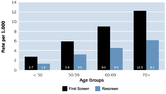
The cancer detection rate was higher among women having their first screen as compared with subsequent screens (Figure 5). The cancer detection rate increased with advancing age for both first screens and rescreens.
As shown in Table 8, few cancers were detected by clinical breast examination alone. Among women 50 and over, the cancer detection rates compare well with standards set by the U.K. and Australia (see Table 3).
| Figure 5 Cancer detection per 1,000 screens by age group, 1996 |
||||||||||||||||||||||||||||||||||||||||||||||||||||||||||||||||||||||||||||||||||||||||||||||||||||||||||||||||||||||||||||||||||
Screening activity in 1996 resulted in the detection of 1,443 breast cancers, 82.7% of which were invasive (Table 9). The remaining 17.3% of cancers were ductal carcinoma in situ (DCIS). DCIS occurs when the cancer cells are located within a milk duct and there is no invasion of the surrounding fatty breast tissue. The proportion of screen-detected cancers that were DCIS declined with increasing age. The overall proportion of in situ cancers (17.3%) lies within the standards developed by the Australian program (10%-20%). It is important that most of the cancers detected are small invasive ones rather than in situ cancers, because the detection of a large number of in situ cancers may not result in a reduction in breast cancer mortality.9 The primary objective of screening is to detect invasive cancers before they have had a chance to spread in the body. This involves detecting breast cancers at a small size (< 15 mm diameter) and without axillary (armpit) lymph node involvement at diagnosis. Stage, tumour size and nodal involvement are three outcomes that allow programs to measure the extent of invasive cancer at the time of diagnosis. In terms of these outcomes, Canadian breast screening programs compare favourably with previously established standards and guidelines. Tabar et al9 state that to achieve a substantial reduction in mortality, 50% of screen-detected invasive cancers should be less than 15 mm in diameter, and at least 70% of screen-detected tumours should not have lymph node metastases. Within Canadian breast screening programs, of invasive cancers detected, 50.8% were less than 15 mm and 77.7% did not have lymph node metastases. The guidelines developed by Tabar et al9 and national programs such as those in the U.K.11 and Australia12 were developed for women in target age groups (i.e. 50-64 or 50-69). As Table 9 shows, outcomes from Canadian provinces are particularly favourable for women between the ages of 50 and 69 years.
|
||||||||||||||||||||||||||||||||||||||||||||||||||||||||||||||||||||||||||||||||||||||||||||||||||||||||||||||||||||||||||||||||||
[Previous] [Table of Contents] [Next]