
Public Health Agency of Canada
www.publichealth.gc.ca
Common menu bar links
Institutional links
Diseases & Conditions
Health & Safety
Research & Statistics
Agency Information
Search Box
E-mail this page
Diagnostic Investigations
Further evaluation of suspicious or uncertain findings following a breast screening examination is a normal part of screening. The success of screening programs in reducing breast cancer mortality in the population depends on the adequacy of follow-up in women with abnormal screens. In 1997 and 1998, complete follow-up information was available for over 90% of women with abnormal screening examinations. Among women screened within organized breast screening programs, 8.1% were referred for additional assessment. For every 100 women with an abnormality found on screening, between six and seven women were subsequently diagnosed with cancer. Those found to be normal are again eligible for routine screening in another 2 years.
To establish or exclude the presence of cancer when a lump or lesion is detected through clinical breast examination or mammography screening, additional assessment is normally required. In Canadian screening programs, women with screen-detected abnormalities and their family physicians are notified by the screening program of the need for further assessment and, for the most part, family physicians coordinate follow-up. Because mammography screening is offered to well women and breast cancer is not present in the majority of women with screening abnormalities, morbidity associated with fear, anxiety and subsequent testing should be minimized by providing a well-coordinated follow-up that assures a firm diagnosis in a timely fashion with the minimum number of interventions.
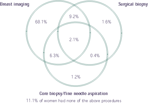
Following an abnormal screening, further investigations may include clinical evaluation, radiologic work-up including diagnostic mammography with additional views, spot compression or magnification views, a comparison with previous mammograms, and ultrasonography. A majority of women aged 50 to 69 (85.7%) underwent some type of imaging procedure, either a diagnostic mammogram and/or ultrasound (Table 5). For 68.1% of women aged 50 to 69, this was the only assessment required. A further 11.1% did not undergo imaging or biopsy, but likely underwent a clinical assessment, and some may have immediately proceeded to a surgical consultation without further intervention (Figure 10).
|
Table 5 |
||||
|
Diagnostic Procedure |
Modes of Detection |
|||
|
All Modes of Detection |
Clinically Detected |
Mammographically Detected |
Mammographically and Clinically Detected |
|
|
Number*(%) |
Number* (%) |
Number* (%) |
Number* (%) |
|
|
Diagnostic mammogram |
30,332 (70.3) |
538 (8.7) |
28,324 (81.8) |
1,470 (62.4) |
|
Ultrasound |
18,532 (42.9) |
1,749 (28.3) |
15,428 (44.6) |
1,355 (57.5) |
|
Fine needle aspiration |
2,173 (5.0) |
533 (8.6) |
1,376 (4.0) |
264 (11.2) |
|
Core biopsy |
2,241 (5.2) |
54 (0.9) |
1,895 (5.5) |
292 (12.4) |
|
Open biopsy with or without fine wire localization |
5,733 (13.3) |
530 (8.6) |
4,654 (13.4) |
549 (23.3) |
|
* All provinces combined. Note: Data for the New Brunswick program are incomplete and therefore do not comprehensively reflect program activity. |
||||
|
Figure 10 |
 |
|
Note: Data for the New Brunswick program are incomplete and
therefore do not comprehensively |
A small number of women may require a surgical consultation, fine-needle aspiration or core biopsy, and surgical biopsy as appropriate to achieve a final diagnosis18,19. More often, less invasive fine-needle aspiration or core biopsy is conducted before resorting to open surgical biopsy. In 1997 and 1998, 17.8% of women received an open surgical biopsy to confirm their diagnosis. Of every 100 women having a surgical biopsy, approximately 38 were found to have cancer. This represents a benign:malignant biopsy ratio of 1.6:1.0, which is within the standards set by other countries (Appendix 1). Keeping the recall rate and the ratio of benign to malignant biopsies appropriately low are important indicators that screening is not inducing unnecessary morbidity in healthy women. Maintaining a low probability of false-positive findings and the resultant invasive procedures has been a challenge in some settings, particularly among women who follow the recommendation for regular screening20.
|
The benign:malignant biopsy ratio of 1.6:1.0 is appropriately low, indicating that screening is not causing unnecessary morbidity in healthy women. |
[Previous] [Table of Contents] [Next]