
Public Health Agency of Canada
www.publichealth.gc.ca
Common menu bar links
Institutional links
Diseases & Conditions
Health & Safety
Research & Statistics
Agency Information
Search Box
E-mail this page
Cancer Detection
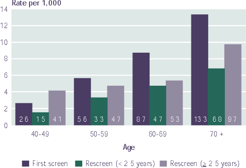
The cancer detection rate increased with age for initial and subsequent program screens (Figure 11). This rate is lower for rescreens occurring less than 2.5 years from the previous screen compared with rescreens occurring at least 2.5 years after the previous screen. This is anticipated as more cancers have the opportunity to develop if the interval between screens is extended. Table 6 shows that 5% to 7% of cancers were detected by clinical breast examination alone. Among women aged 50 and over, the cancer detection rates measure up well with the standards set by the UK and Australia (Appendix 1).
A total of 3,975 cancers were detected for the screen years 1997 and 1998, of which 80.2% were invasive and 19.8% were ductal carcinoma in situ (DCIS) (Table 7). The proportion of screen-detected cancers that were invasive increased with age. The overall proportion of in situ cancers (19.8%) is within Australian standards (10% to 20%).
|
Figure 11 |
 |
|
Notes: Quebec data are not included. Data for the New Brunswick program are incomplete and therefore do not comprehensively reflect program activity. |
|
Preventing breast cancer deaths through mammographic screening depends on detecting cancers early, before they can be felt: 37.6% of invasive cancers were detected at <= 10 mm diameter and 78.5% were lymph node negative. |
|
Table 6 |
|||||
|
Mode of Detection |
40-49 |
50-59 |
60-69 |
70+ |
All Ages |
|
Detected by mammography alone |
|
|
|
|
|
|
Detected by both mammography and CBE* |
|
|
|
|
|
|
Detected by CBE alone* |
|
|
|
|
|
|
All modes of detection |
|
|
|
|
|
|
* Manitoba, Ontario, Nova Scotia and Newfoundland provide CBE by a nurse or technologist; of these programs, all but Nova Scotia restrict program participation to women aged 50 and older. Notes: The Quebec program has incomplete cancer information due to incomplete data linkages. Therefore, Quebec data are excluded from this table. Data for the New Brunswick program are incomplete and therefore do not comprehensively reflect program activity. |
|||||
The secondary prevention of breast cancer death through mammographic screening depends on detecting cancers at an early stage, before they can be felt, leading to more treatment options, reduced recurrence and improved survival21. Nearly 90% of women with stage I cancers survive at least 5 years; this stage accounted for 50.9% of screen-detected cancers in women aged 50 to 69. Survival decreases as the stage of the cancer increases, reflecting larger tumours and more lymph node involvement. Five year survival rates are 75% for women with stage II cancers, just over 40% for stage III, and just under 20% for stage IV cancers2.
The Europe Against Cancer guidelines recommend that to achieve a substantial reduction in mortality, 25% or more of screen-detected invasive cancers should be <= 10 mm in diameter. Swedish standards also recommend that at least 70% of screen-detected tumours should not have lymph node metastases. Once again, Canadian breast screening programs fared well as 37.6% of invasive cancers were detected at <= 10 mm diameter and 78.5% were lymph node negative (Table 8).
Even though abnormal recall rates did not differ with age (Table 4), the positive predictive value (PPV) increased with age (Figure 12), reflecting the increased number of cancers with advancing age and improved discriminating power of mammograms for less dense breasts. Delayed (>= 2.5 years) intervals to rescreen tended to increase cancer detection rates. Within age groups, PPVs were similar on first and subsequent screens, but increased with age. This may reflect the fact that PPV values increase as the prevalence of cancer increases.
|
Table 7 |
||||||||||
|
40-49 |
50-59 |
60-69 |
70+ |
All Ages |
||||||
|
n |
% |
n |
% |
n |
% |
n |
% |
n |
% |
|
|
Number of cancers |
|
|
|
|
|
|
|
|
|
|
|
TNM staging |
|
|
|
|
|
|
|
|
|
|
|
Tumour size (invasive only) |
|
|
|
|
|
|
|
|
|
|
|
Positive nodes (invasive only) |
|
|
|
|
|
|
|
|
|
|
|
* Includes missing values and cases in which dissection was not done. Notes: The Quebec program has incomplete cancer information due to incomplete data linkages. Therefore, Quebec data are excluded from this table. Data for the New Brunswick program are incomplete and therefore do not comprehensively reflect program activity. |
||||||||||
[Previous] [Table of Contents] [Next]